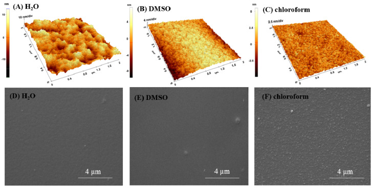
Figure 4.
AFM surface images (3D-up) and SEM micrographs (down) on dry layers of p(NIPAM-BA) deposited by spin coating on Si with different rotational parameters (rpm) for each solvent: water, 4500/20 s (A,D); DMSO × 2 steps (5000/1 min 30 s and 6000/50 s) (B,E); chloroform, 4500/10 s (C,F). The AFM images correspond to areas of 5 × 5 µm2 and 2 × 2 µm2. SEM magnifications 20,000×.

