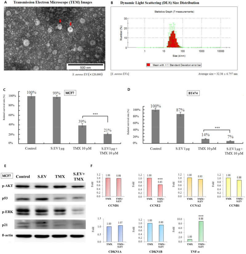
Figure 4.
Effect of combination treatment with S. aureus EVs and tamoxifen in breast cancer cells. (A) EVs derived from S. aureus (indicated by a red arrow) were confirmed by transmission electron microscopy. (B) The average diameter of S. aureus EVs was obtained using dynamic light scattering size distribution. (C) The bar chart shows the percentage of viable MCF7 cells after treatment with S. aureus EVs and/or tamoxifen. (D) Relative BT474 cell survival percentages after treatment with S. aureus EVs and/or tamoxifen. This experiment was repeated three times. (E) Protein expression of p−AKT, p53, p−ERK, and p21 was detected by Western blotting after tamoxifen and/or S. aureus EVs treatment. lane 1, control; lane 2, S. aureus EVs 100 ng/mL; lane 3, 10 μM tamoxifen; lane 4, 10 μM tamoxifen plus S. aureus EVs 100 ng/mL. (F) The mRNA expression of cyclins, cyclin−dependent kinase inhibitors, and TNF−α was confirmed by qRT−PCR. CCND1, cyclin D1; CCNE2, cyclin E2; CCNA2, cyclin A2; CCNB1, cyclin B1, CDKN1A:p21, and CDKN1B:p27; TNF−α, tumor necrosis factor−α. *** p < 0.001.

