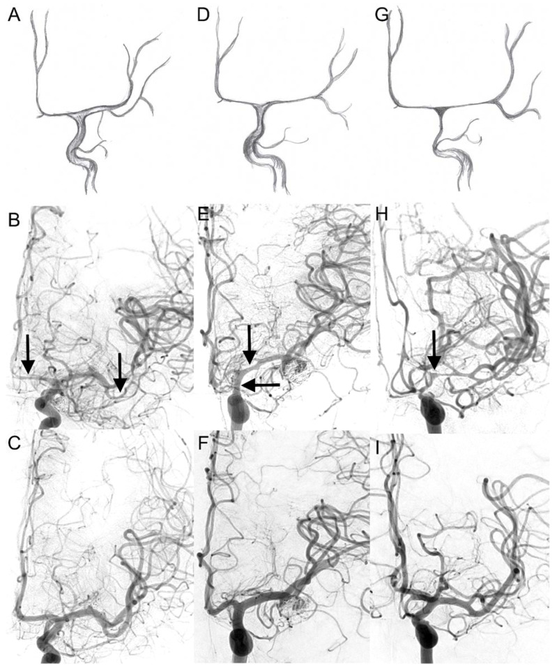
Figure 1.
Visual cerebral vasospasm classification in digital subtraction angiography. The upper two lines display cerebral vasospasm grades (CVSG) as drawings with the corresponding angiograms. In the bottom line, the corresponding control DSA after six months is shown: (A,B) CVSG 1—Narrowing of the A2, A1, and M2 segments (arrows) with postspastic enlargement of distal M2 branches. (D,E) CVSG 2—The spasm affects the proximal M1 segment and the intradural carotid artery (arrows). (G,H) CVSG 3—The intradural carotid artery, the proximal middle cerebral artery, and the anterior cerebral artery show high-grade narrowing with a fading appearance like a ghost (arrow). (C,F,I)—control DSA after 6 months (CVSG 0).

