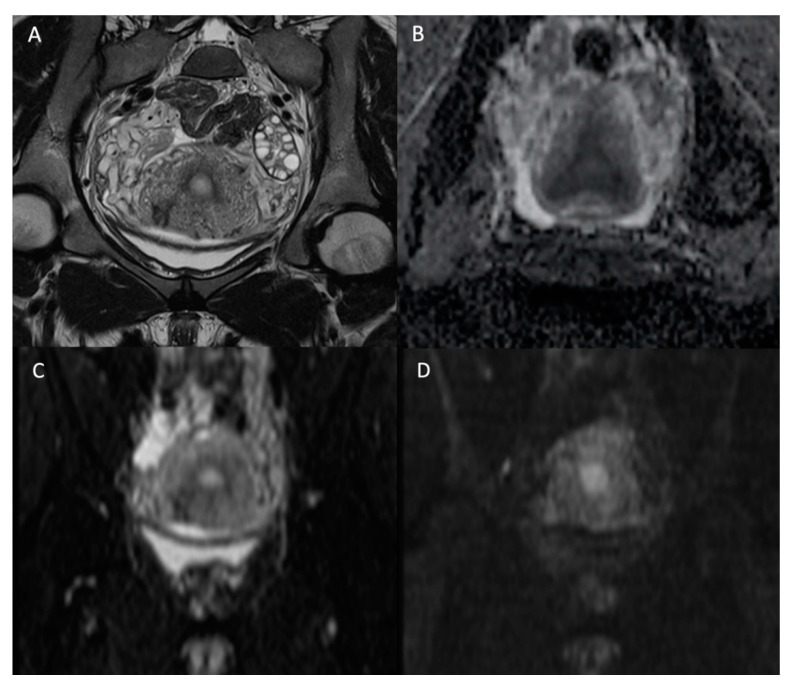
Figure 1.
Normal female pelvis of 26-year-old in the coronal plane. (A) T2W image; (B) ADC map; (C) b-value = 0 s/mm2 DW image; (D) b-value = 1000 s/mm2 DW image. We see the disappearance of high fluid signal (as the one in the bladder) with increasing b-values but persistence of high signal intensity on high b-value for the endometrium.

