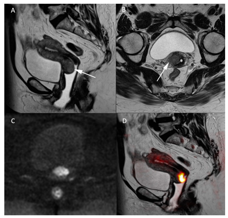
Figure 2.
MR images of a 66-year-old woman with a known cervical carcinoma. (A) Sagittal T2W image; (B) axial T2W image perpendicular to the cervical axis. Cervical cancer and its extension appearing as low-contrasted T2W area (arrow) through the normal stroma and the right parametrium, (C) high b-value (b = 1000 s/mm2) and (D) fusion images between T2W and high b-value sequences for better evaluation of the carcinoma’s extension.

