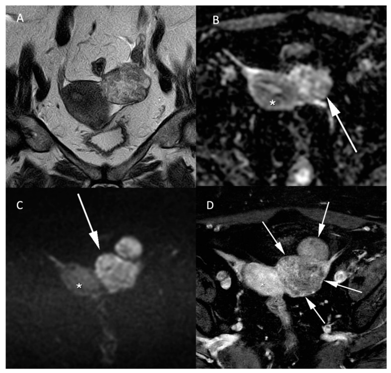
Figure 5.
Histologically proven left ovary adenocarcinoma in a 64-year-old woman. (A) T2W hyperintense heterogeneous left adnexal mass next to the uterus (*). Tissular bilobed left adnexal mass with parts of low (B) ADC values and high (C) b-1000 signal consistent with a diffusion restriction in the lesion (C). Post injection of gadolinium (D) T1W sequence with fat-saturation shows a heterogeneous enhancement (arrow).

