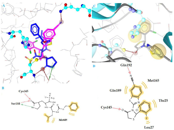
Figure 3.
(A) Superposition of compound k3 (magenta) and inhibitor GC376 (blue) bound to SARS-CoV-2 main protease structure 6M2N, with specific residues labeled. (B) 2D interaction diagram of inhibitor GC376 docking pose interactions with the key amino acids. (C) Docking pose of compound k3 in SARS-CoV-2 main protease structure 6M2N. (D) 2D interaction diagram of compound k3 docking pose interactions with the key amino acids. Red and green dotted arrows indicate H-bond and yellow spheres hydrophobic interactions.

