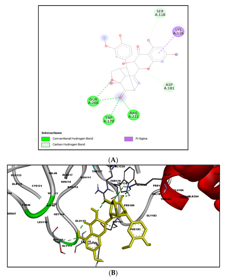
Figure 2.
Molecular interactions of silydianin with the catalytic active site of PTP1B protein in Schrödinger’s suite. (A) Two-dimensional view. (B) Three-dimensional view. The dashed lines represent the bond formations between the ligand and amino acids (protein). Trp179, Arg221 and Gln266 form conventional H-bonds (dark green color), Ser118 and Asp181 form carbon–hydrogen bonds (light green color), while Lys116 forms Pi-sigma bonds (purple color).

