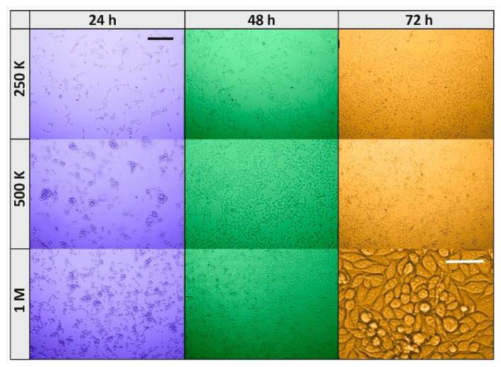
Figure 9.
Microphotograph of RKO cells plated at various times of withdrawal from the incubator and at different densities. Cells plated at 250 K, 500 K and 1 M cells/dish withdrawn from the incubator after 24 h (blue background), 48 h (green), and 72 h (orange); black scale bar is 200, and it applies to all microphotographs, but the white scale bar (50 µm) applies to its microphotograph only.

