. 2022 Mar 31;11:e71469. doi: 10.7554/eLife.71469

Figure 5. Lar and Sns expression in the developing pupal mushroom body (MB).

Confocal projections and single optical slices showing Lar and Sns expression in the 24 hr after puparium formation (APF) (A–D’) and 40 hr APF (E–H’) pupal MB, co-stained with FasII antibody (magenta). Projections of the entire MB are shown in (A, A’, C, C’, E, E’, G, and G’). The rest are single optical slices. Lar expression is seen in the growth cones of developing β lobe axons (B, B’, arrows). No Sns expression is seen in the 24 hr APF MB (C–D’). Sns expression is seen in neuronal projections at the midline (D’, arrow). (E–F’) Lar expression is seen in α, α′, β, and β′ lobes in the 40 hr APF MB (arrows in E’; F, F’, single slice showing all four lobes with Lar expression). (G–H’) No Sns expression is seen in the 40 hr APF MB (asterisks in H’ denote unlabeled α, α′, β, and β′ lobes). Scale bars, 20 µm. See Figure 5—figure supplement 1 for Lar and Sns expression in 72 hr APF and adult brains and Figure 5—figure supplement 2 for Lar and Sns expression in the pupal and adult antennal lobes.

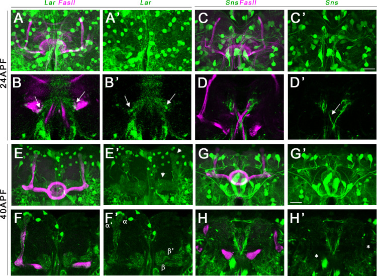
Figure 5.

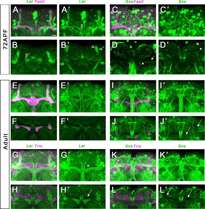
Figure 5—figure supplement 1. Lar and Sns expression in the 72 hr after puparium formation (APF) and adult mushroom body (MB).

Figure 5—figure supplement 1.

Confocal projections and single optical slices showing Lar and Sns expression in the 72 hr APF (A–D’) and adult (E–L’) MB, co-stained with FasII antibody (magenta). Projections of the entire MB are shown in A, A’, C, C’, E, E’, G, G’, I, I’, K, and K’. The rest are single optical slices. (A–B’) Strong Lar expression is seen in all lobes of the 72 hr APF MB. (C–D’) Weak Sns expression is seen in α and β lobes of the 72 hr APF MB (D–D’, arrows). Expression is seen only in older axons in the α and β lobes with hollow cores in the center of the lobes (D–D’, asterisks). Lar continues to be strongly expressed in α and β lobes of the adult MB (E–H’). (F–F’) Single optical slice from the adult MB showing strong Lar>GFP expression in the α and β lobes, co-localizing with FasII staining. (G, G’) Projection of confocal slices showing the entire adult MB, double-stained for GFP (green) and Trio (magenta). Trio labels the γ, α′, and β′ lobes of the adult MB. (H, H’) Single optical slice showing no detectable Lar>GFP expression in the Trio-expressing α′ and β′ lobes (H’, arrow). Weak expression is seen in the γ lobe (G’, asterisk). (J, J’) Single optical slice showing faint Sns expression in the α and β lobes (J’, arrows). (L, L’) Single optical slice showing no Sns expression in the γ, α′, and β′ lobes (L’, arrows).
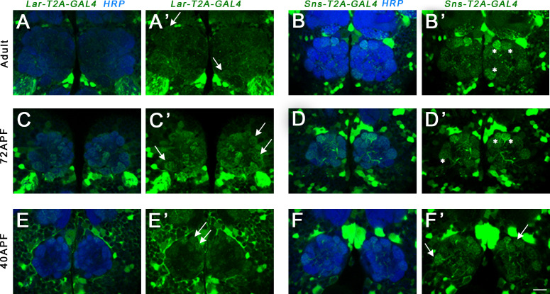
Figure 5—figure supplement 2. Lar and Sns expression in the adult and developing pupal antennal lobes (ALs).

Figure 5—figure supplement 2.

Single optical slices showing either Lar>GFP or Sns>GFP (green) expression in the ALs, co-stained with anti-HRP (blue) to label the AL neuropil. (A, A’) Weak Lar>GFP expression seen in several glomeruli of the adult AL (A’, arrows). (B, B’) Strong Sns>GFP expression seen in three glomeruli of the adult AL (B’, asterisks). (C, C’) Strong Lar>GFP expression seen in several glomeruli in the 72 hr after puparium formation (APF) AL (C’, arrows). (D, D’) Sns>GFP is expressed at high levels in some glomeruli in the 72 hr APF AL (D’, asterisks). (E, E’) Weak Lar>GFP expression is seen in several glomeruli in the 40 hr APF AL (E’, arrows). (F, F’) Sns>GFP is strongly expressed in several glomeruli in the 40 hr APF AL (F’, arrows). Scale bar, 20 µm.