. 2022 Mar 31;11:e71469. doi: 10.7554/eLife.71469

Figure 7. Lar and Sns expression in the developing pupal optic lobes (OLs).

Single optical slices showing Lar and Sns (green) expression in 24 hr after puparium formation (APF), 40 hr APF, and 72 hr APF OL, co-stained with anti-Chaoptin (24B10 mAb, red). (A, A’) Weak Lar>GFP expression is seen in the medulla neuropil at 24 hr APF. (B, B’) At this timepoint, Sns>GFP is expressed at high levels in neuronal cell bodies in the medullary cortex (B’, arrow) and in specific layers in the medulla and lobula. (C, C’) Strong Lar>GFP expression is seen in L1 lamina neuron cell bodies (C’, arrow), which arborize in layers M1 and M5 of the medulla in the 40 hr APF OL. (D–D’’) Close-up of the distal medulla showing L1 lamina neuron arbors in M1 and M5 layers of the medulla (D’, asterisks). Faint Lar expression is seen in the M6 layer of the medulla (D’’, arrowhead). (E, E’) Sns>GFP expression increases at 40 hr APF, with many more neurons expressing Sns in the medullary cortex. Sns>GFP expression can be seen in several layers in the distal as well as the proximal medulla. (F–F’’) Close-up of the distal medulla showing Sns>GFP expression in layers M1 through M5 of the medulla. Sns is not expressed in R7 photoreceptors. (G–H’’) Strong Lar>GFP expression seen in L1 cell bodies (G’, arrow) and layers M1 and M5 of the medulla (H’, asterisks). Strong Lar expression is seen in close proximity to R7 axons and terminals (H, H’, arrows; H’’, arrowhead). (I–J’’) Sns>GFP is expressed at very high levels in the 72 hr APF OL. Specific Sns expression is seen in M1, M5, and M10 layers of the medulla and a few layers in the lobula (I, I’). Sns expression is also seen in the lamina. Note the dots at the top of the lamina (arrows in E’ and I’), which match the morphologies of C2 endings. C2 arborizes in layers M1, M5, and M10 of the medulla. (J–J’’) Close-up of the distal medulla showing Sns expression in M1 and M5 layers. Scale bar, 20 µm. See Figure 7—figure supplement 1 for Lar and Sns expression in the adult OL and Sns expression in a C2-like arborization pattern. See Figure 7—figure supplement 2 for further characterization of Lar and Sns OL expression.

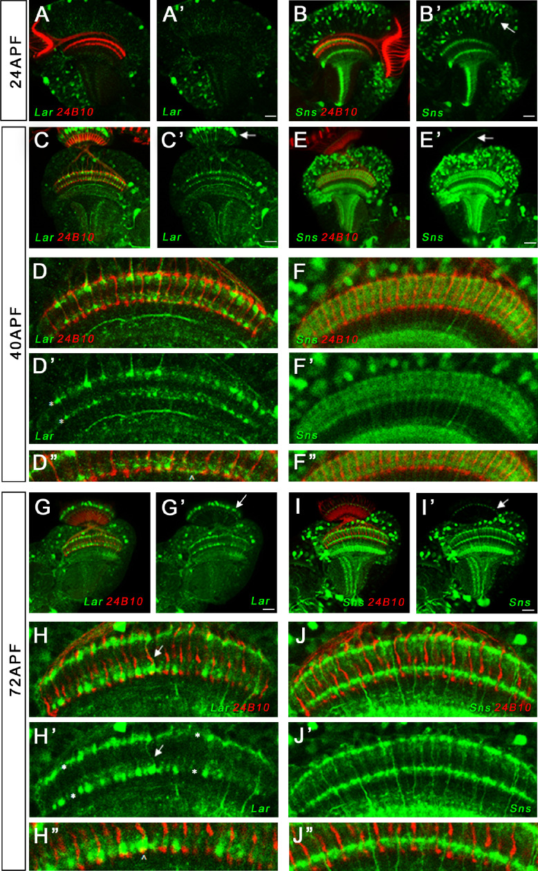
Figure 7.

Figure 7—figure supplement 1. Expression of Lar and Sns in the adult optic lobe (OL).

Figure 7—figure supplement 1.

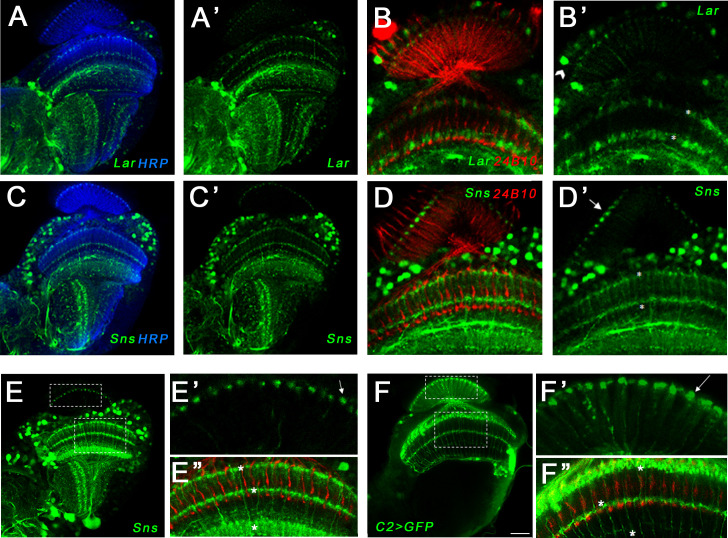
Confocal projections and single optical slices showing Lar>GFP and Sns>GFP expression in the adult OL. Brains were double-stained for GFP (green) and either anti-HRP (blue) or Chaoptin (24B10; red), which labels photoreceptor axons. (A, A’) Single optical slice showing Lar expression (green) in the adult fly OL. (B, B’) Single optical slice showing Lar>GFP expression in L1 Lamina neurons (B’, arrowhead) and in layers M1 and M5 (asterisks in B’) of the adult medulla. (C–D’) Single optical slices showing Sns>GFP expression (green) in the adult OL. Strong Sns>GFP expression is seen in layers M1 and M5 of the adult medulla (D’, asterisks). Sns expression is also seen in the same dot-like pattern at the top of the lamina neuropil as seen in the pupal OL (D’, arrow). Sns>GFP does not appear to colocalize with 24B10, suggesting that it is not in R7 or R8 photoreceptors. (E–E’’) Single optical slice showing Sns expression in the 72 hr after puparium formation (APF) OL. Entire OL is shown in (E). (E’) shows close-up of the lamina. Note the dot-like endings of Sns-expressing neurons in the lamina neuropil. These endings most closely resemble those of C2 neurons (Tuthill et al., 2013). (E’’) Sns reporter expression seen in M1, M5, and M10 layers of the medulla (asterisks), resembling C2 arborization pattern in the medulla (Tuthill et al., 2013). (F–F’’) Single optical slice showing the projection pattern of C2 neurons in the adult OL. A split-GAL4 driver (Tuthill et al., 2013) was crossed to UAS-GFP reporter to visualize the arborization pattern. The dot-like endings at the top of the lamina neuropil (F’, arrow) are similar to the dot-like endings seen with Sns in (E’). C2 neurons arborize in M1, M5, and M10 layers of the medulla (F’’, asterisks), similar to Sns (E’’). Scale bar, 20 µm.
Figure 7—figure supplement 2. Dendritic projections of Sns neurons in the adult optic lobe (OL), Lar>GFP expression in the 24 hr after puparium formation (APF) retina and LarMiMIC expression in L1 lamina neurons.

Figure 7—figure supplement 2.

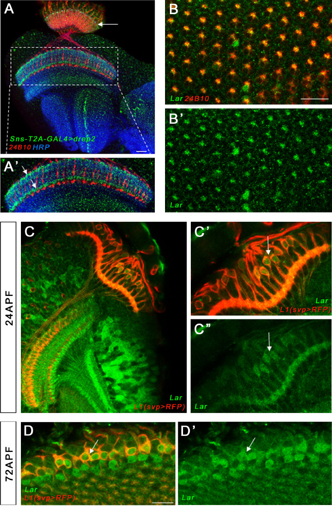
(A, A’) Single optical slice showing dendritic projections of Sns-expressing neurons visualized by Sns-GAL4>Drep2 (dendrite-specific marker, green), co-stained with 24B10 (red) and anti-HRP (blue). Specific dendritic projections can be seen in M1 and M5 layers of the medulla (A’, arrows) and at the top of the lamina neuropil (A, arrow). C2 is known to receive synapses in M1, M5, and the lamina. A single layer is seen in the lobula. (B, B’) Lar expression is seen in a top-down view of the 24 hr APF retina, co-stained with 24B10, (red). R7 and R8 photoreceptor cell bodies lie in the center of each ommatidium. Lar expression is seen in R7 and/or R8 cell bodies in the center of the ommatidia as well as in at least one outer photoreceptor (R1–R6). Single optical slice focused on the central R7/R8 photoreceptors is shown. (C–D’) LarMIMIC expression was visualized by GFP immunostaining. We used an L1 Lamina neuron-specific driver (svp-GAL4) and UAS-mCD8-RFP reporter to visualize L1 neurons in the pupal OL. LarMiMIC co-localized with RFP-expressing L1 lamina neurons in the 24 hr APF and 72 hr APF pupal lamina (C’–D’, arrows). Scale bar, 20 µm.