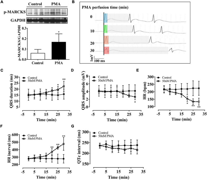
FIGURE 1.
Phorbol-12-myristate-13-acetate (PMA) perfusion induced prolongation and low amplitude of the QRS complex in isolated normal rat hearts. The control hearts were perfused with the Krebs–Henseleit (KH) buffer solution. (A) Western blot analysis showing that PMA perfusion (50 nm) activated phospho-myristoylated alanine-rich C kinase substrate (MARCKS) protein. (B) Representative ECG recording in lead II showed QRS prolongation of the isolated hearts after PMA perfusion for 0, 10, 20, and 30 min. Light blue box: QRS duration of isolated heart after stabilization; light green box: QRS duration of isolated heart after 10-min of perfusion; light orange box: QRS duration of isolated heart after 20-min of perfusion; light red box: QRS duration of isolated heart after 30-min of perfusion; light gray box: RR interval. (C–G) Statistical data of electrocardiography (ECG), QRS duration (C), QRS amplitude (D), hear rate (HR) (E), RR interval (F), and QTc interval (G). Error bars represent mean ± SD, n = 5 for each group. *p < 0.05 vs. control, **p < 0.01 vs. control.

