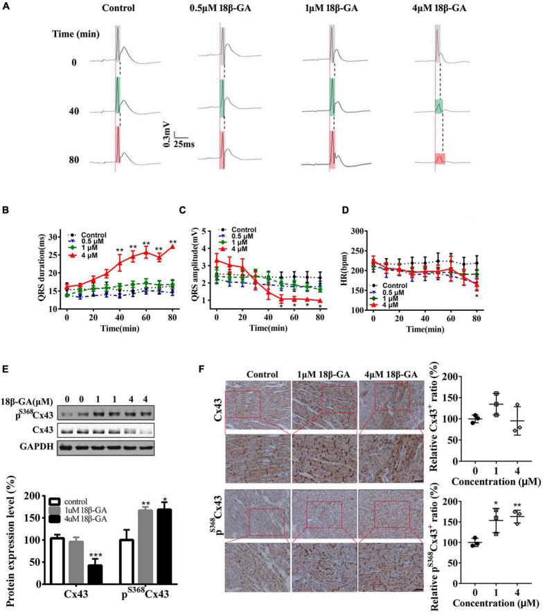
FIGURE 3.
Cx43 hyperphosphorylation and lateralization induced by 18β-GA produce an abnormal QRS complex. (A) 18β-GA (0.5, 1, and 4 μm) perfusion (80 min) produced abnormal QRS duration of isolated rat hearts. Light gray box: QRS duration of isolated heart after stabilization; light green box: QRS duration of isolated heart after 40-min of perfusion; light red box: QRS duration of isolated heart after 80-min of perfusion. (B–D) Statistical data changes of QRS duration, QRS amplitude, and HR. n = 5 for each group. (E) Western blot images of pS368Cx43 and Cx43 in the control and 18β-GA-treated rats’ cardiac ventricles. The level of pS368Cx43 and Cx43 was normalized to GAPDH. (F) Immunohistochemical analysis of Cx43 and pS368Cx43 expression and redistribution (Bar = 20 μm), and corresponding statistical data of Cx43+ and pS368Cx43+ cells ratio. Error bars represent mean ± SD. *p < 0.05 vs. control; **p < 0.01 vs. control; ***p < 0.001 vs. control.

