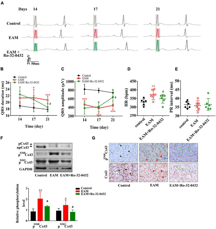
FIGURE 4.
Blockade of protein kinase C (PKC) with Ro-32-0432 prevented the prolonged duration and low voltage of the QRS complex by suppressing Cx43 hyperphosphorylation and redistribution. (A) Representative ECG recording of normal, experimental autoimmune myocarditis (EAM), and Ro-32-0432-treated EAM rats. Each set of the ECG was recorded from the same rat on days 14, 17, and 21. Light gray box: QRS duration of control rats; light red box: QRS duration of EAM rats; light green box: QRS duration of Ro-32-0432-treated EAM rats. (B–E) Statistical data of ECG, QRS duration (B), QRS amplitude (C), HR (D), and PR interval (E). (F) Western blot images of pS368Cx43, pS262Cx43, and Cx43 in the cardiac ventricles of control, EAM, and Ro-32-0432-treated EAM rats. n = 6 for each group. (G) Immunohistochemical analysis of Cx43 and pS368Cx43 expression and redistribution (Bar = 20 μm). The black arrows in the pictures of the control group indicate the normal pattern of Cx43 and pS368Cx43 in intercalated disc; the red arrows indicate the lateralization of Cx43 and pS368Cx43 in the EAM group; the green arrows indicate partially recovered pattern and distribution of Cx43 and pS368Cx43 in Ro-32-0432-treated EAM group. Error bars represent mean ± SD. *p < 0.05 vs. control; **p < 0.01 vs. control; ***p < 0.001 vs. control; #p < 0.05 vs. EAM.

