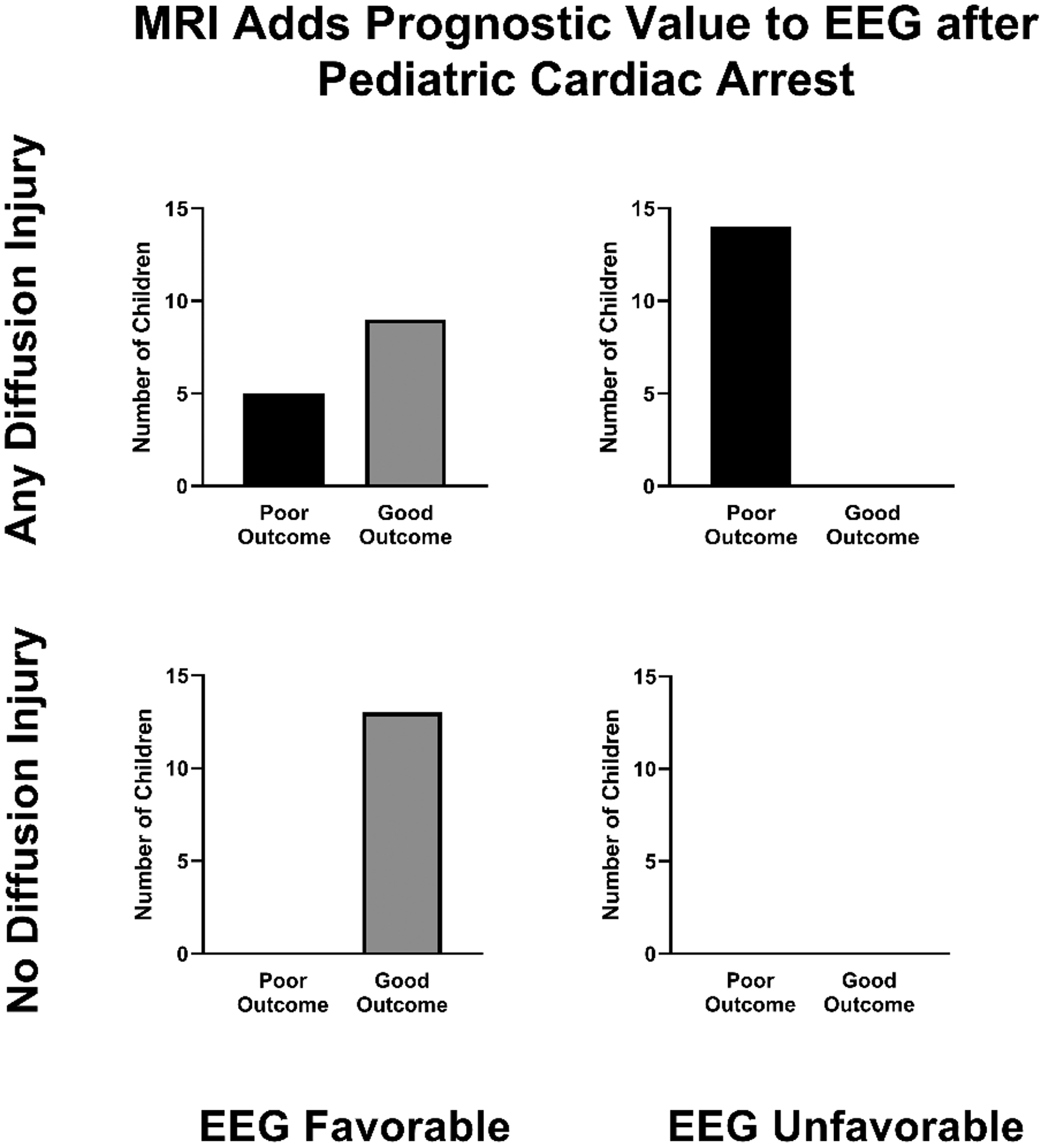
Fig. 3:

MRI diffusion abnormalities with EEG background in relation to outcome at hospital discharge. All children with poor outcome had MRI diffusion abnormalities, of which more had an unfavorable EEG background than favorable. No child with unfavorable EEG background had normal diffusion imaging. Favorable EEG background includes normal or slow-disorganized and unfavorable EEG background includes discontinuous/burst-suppression or attenuated-featureless. EEG=electroencephalogram; MRI= magnetic resonance imaging.
