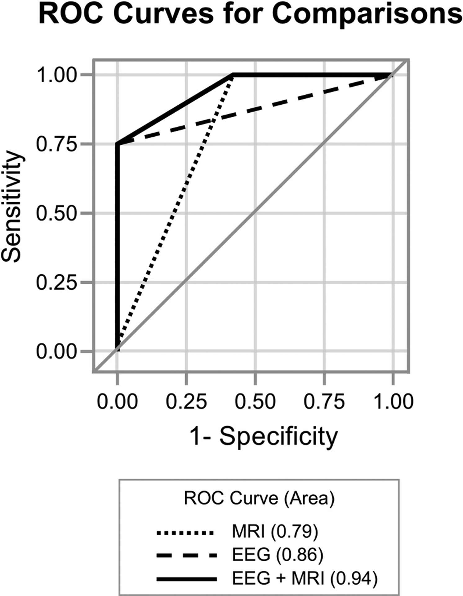
Fig. 4. Multimodal test results improve ability to discriminate poor outcome after pediatric cardiac arrest.

ROC curves depicting EEG suppressed background (Category 3 or 4, AUC=0.86), MRI diffusion abnormalities (AUC=0.79) and combined (EEG+MRI, AUC=0.94) were compared. A model combining EEG suppressed background with MRI diffusion abnormalities is better able to distinguish poor outcome than either EEG (p=0.02) or MRI alone (p=0.0008). EEG=electroencephalogram performed less than 72 hours after cardiac arrest; MRI=magnetic resonance imaging, post-arrest; AUC=area under the ROC curve.
