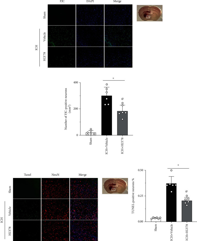
Figure 5.

Effects of HLY78 on neuronal damage at 24 h after ICH. (a) Representative micrographs of FJC staining in the perihematomal area, and (b) quantitative analysis of FJC-positive neurons 24 h after ICH. (c) Representative micrographs of TUNEL staining in the perihematomal area, and (d) quantitative analysis of TUNEL-positive neurons. 24 h after ICH. Scale bar = 100 μm. ∗p < 0.05 vs. sham group; @p < 0.05 vs. ICH+vehicle group. One-way ANOVA and Tukey test, n = 6/group.
