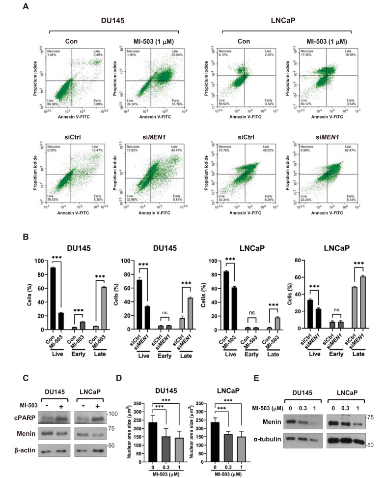
Fig. 2. Inhibition of menin induces apoptosis in PCa cells.
(A and B) FACS analysis of DU145 and LNCaP cells treated with 1 μM of MI-503 (48 h) or siRNAs (48 h). Cells were stained with Annexin V and PI and analyzed by FACS. n = 3, ***P < 0.001; ns, not significant. Con, control. (C) Western blot analysis of DU145 and LNCaP cells treated with MI-503 (1 μM) using specific antibodies as indicated. β-actin was used as a loading control. (D) DAPI staining of MI-503-treated PCa cells. The nuclei of DU145 (n = 50) and LNCaP (n = 61) cells after treatment with MI-503 were DAPI-stained to measure the size of the nuclei. ***P < 0.001. (E) Western blot assay of menin in DU145 and LNCaP cells treated with 0.3 μM or 1 μM of MI-503, respectively. α-tubulin was used as a loading control.

