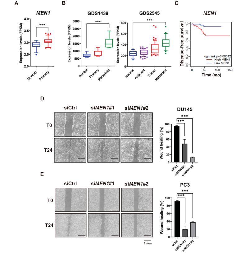
Fig. 4. Menin is highly represented in metastatic cancer and linked to cell migration.
(A) The relative expression of MEN1 in the TCGA cohort (normal = 52, primary prostate tumor = 497). ***P < 0.001. (B) MEN1 expression profiles from the GEO datasets. Raw values were obtained from GDS1439 (benign n = 6, primary n = 7, and metastatic n = 6) and GDS2545 (normal n = 18, adjacent n = 63, tumor n = 65, and metastatic n = 25). ***P < 0.001. (C) Kaplan–Meier disease-free survival analysis of patients with prostate adenocarcinoma and high (n = 156, red line) or low (n = 176, blue line) MEN1 expression. P values were determined using the log-rank test. (D and E) DU145 (D) and PC3 (E) cells were scratched 48 h after siMEN1 transfection and the migration efficiency was analyzed 24 h after scratching. The percentage of wound healing was defined as the extent of relative wound closure (n = 3). Scale bars = 1 mm. ***P < 0.001.

