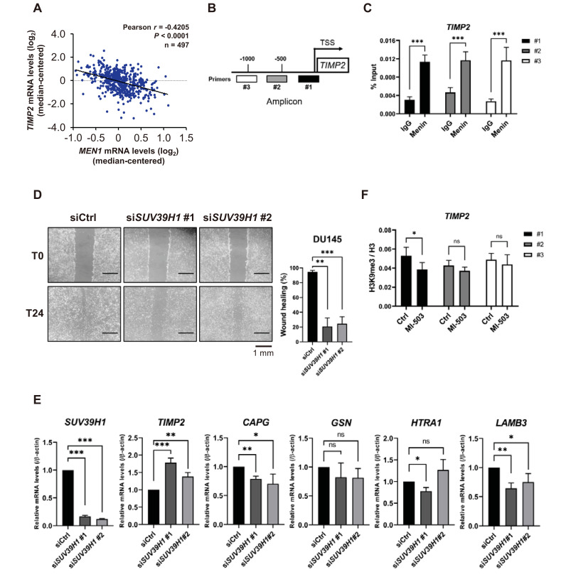
Fig. 6. Menin downregulates TIMP2 and plays a unique role in PCa cell metastasis.
(A) Pearson’s correlation analysis between MEN1 and TIMP2 mRNA levels in prostate primary tumor tissues (n = 497). (B) A diagram of the TIMP2 promoter, showing the locations of ChIP primers. TSS, transcription start site. (C) ChIP was performed using a chromatin solution prepared from DU145 cells treated with either rabbit IgG or anti-menin antibody and analyzed by RT-qPCR with the indicated ChIP primers. Results are shown as the percentage (%) input. ***P < 0.001. (D) DU145 cells were scratched 48 h after siSUV39H1 transfection and the migration efficiency was analyzed 24 h after scratching. The percentage of wound healing was defined as the extent of relative wound closure (n = 3). Scale bars = 1 mm. **P < 0.01; ***P < 0.001. (E) The mRNA level of metastasis-related genes upon SUV39H1 knockdown in DU145 cells. Bars show the mean mRNA level and SD. *P < 0.05; **P < 0.01; ***P < 0.001; ns, not significant. (F) DU145 cells were treated with 1 μM of MI-503 for 48 h. ChIP was performed using rabbit IgG, anti-H3, or anti-H3K9 me3 antibody. H3K9 me3 enrichment was normalized by H3 enrichment. *P < 0.05; ns, not significant.

