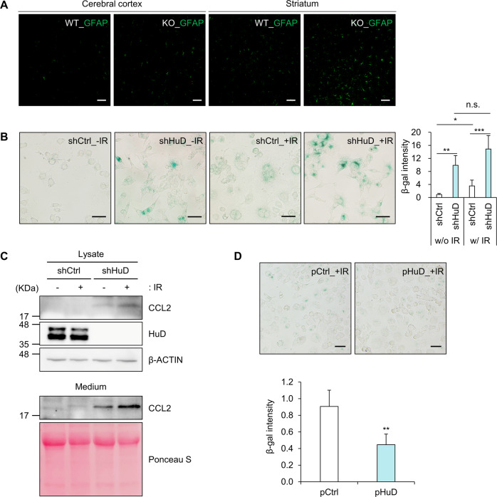
Fig. 5. Sensitization of N2a cells via HuD knockdown in response to senescence inducer.
A Relative level of GFAP, an astrocyte marker, was analyzed in the cerebral cortex and striatum regions in the brain of HuD KO mice and their age-matched control mice by immunofluorescence microscopy. n = 3. B, C After exposure to γ-irradiation (5.5 Gy), the levels of SA β-gal (B) and CCL2 expression (C) were analyzed by β-gal staining and western blotting analysis. D N2a cells were transiently transfected with plasmids and exposed to γ-irradiation. The levels of SA β-gal were assessed by β-gal staining and densitometric analysis using Image J. Scale bar, 50 μm. Images are representative, and data indicate the mean ± SEM derived from three independent experiments. The statistical significance of the data was analyzed via Student’s t-test; *p < 0.05; **p < 0.01; ***p < 0.001; n.s., not significant.

