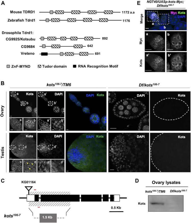
FIGURE 1.
Drosophila CG9925/Kotsubu localizes to the nuclear periphery in germline cells. (A) Structural diagram depicting protein domains of Tdrd1 orthologs in mice, zebra fish, and flies. MYND-type zinc finger domain and Tudor domains are in light gray and stripes, respectively. In fly, an additional N-terminal RNA recognition motif (black) is found in Vreteno but is absent in the other orthologous proteins. (B) Kots is observed as perinuclear foci in germline cells in the germaria (a) and localizes to the nuclear peripheral nuage in the egg chambers (b) in ovaries. In testes, Kots appears as perinuclear foci in the spermatogonia (c) and condenses into enlarged foci (yellow arrowheads) in the spermatocytes (d). Endogenous Kots is absent in deficiency (Df) over kots mutant gonads (Df/kots 106-7 ). (C) Loss-of-function allele of kots was generated by imprecise excision of a P-element insertion, KG01164. Red bars represent the primers used to detect 118 bp of the first exon. (D) Western blotting of ovarian lysates showing a single band for Kots in the heterozygous control that is absent in Df/kots 106-7 mutants. (E) Immunostaining of kots mutant ovaries expressing C-terminal Myc-tagged Kots (Kots-Myc) in germline cells with anti-Myc and Kots antibodies. NGT40 was used to drive the expression of UASp-kots-Myc in the germline. Perinuclear puncta in the germaria (a, a’) and egg chambers (b, b’) are discernible. All scale bars are 10 μm.

