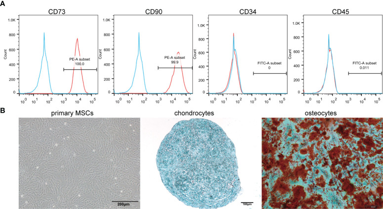
Figure 1.
Identification of MSCs. (A) Immunophenotypic characterization of hMSCs was performed by flow cytometry. The vast majority of cells (>99%) were positive for CD73 and CD90, but a few cells (<0.1%) expressed CD34 and CD45. (B) Primary cells presented as morphologically homogeneous, with elongated spindle appearance. The hMSCs were cultured in appropriate differentiation media for 3 weeks. For chondrogenic differentiation, the fixed chondrospheres were embedded and cut into sections and stained with alcian blue. The induced cells were stained by oil red O to indicate osteocytes.

