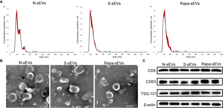
Figure 2.
Identification of sEVs. (A) NTA displayed that the diameters of most sEVs in each group were around 100nm. (B) TEM showed that sEVs in each group were 50–200 nm disc-like vesicles with bilayer membrane. (C) Western blot analysis showed that sEVs of all groups express CD9, CD63, and TSG101.

