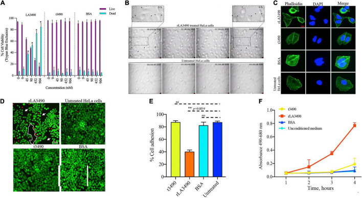
FIGURE 4.
Cytopathic effect of rLA3490. (A) Dose-dependent HeLa cell death induced by r3490 as assessed by trypan blue dye exclusion. Negative controls, t3490, BSA, and no treatment, had no such effect. Cell monolayers were treated with graded molar ratio doses (0 to 904 nM) of LA3490, t3490, and BSA for 4 h. Data represent mean ± SD of two independent experiments done with each condition in triplicate (paired t-test, *p < 0.005). (B) Time-lapse phase-contrast microscopy images (40 frames, 5 s intervals) showing HeLa cell cytopathic effect following exposure to 45 nM of rLA3490 and controls. Only with rLA3490 was cell blebbing evident from 1 h onward [seen in zoom view (top left and right panel, black arrow)]. Time-lapse imaging was captured using a × 40 objective lens using a Leica DMi8 inverted microscope. Scale bar, 10 μm. (C) Actin depolymerization occurs early after rLA3490 treatment. HeLa cell monolayers were incubated with 45 nM of rLA3490, t3490, and BSA up to 1 h. Monolayers were fixed with 4% paraformaldehyde followed by 0.1% Triton X-100 in PBS permeabilization. The monolayer was incubated with phalloidin-Alexafluor-488 nm conjugate, washed, and then mounted with ProLong™ Gold Antifade Mountant with DAPI. Images were captured using a Leica DMi8 confocal microscope [Alexa_488 nm (green), DAPI (blue)] at × 40 magnification. Untreated HeLa cells served as control. Scale bar, 20 μm. (D) HeLa cell death induced by rLA3490 as assessed by fluorescent live/dead staining. Negative controls (t3490, BSA, and no treatment) had no such effect. Live/dead staining of HeLa cell monolayers was carried out after 4-h exposure to 45 nM rLA3490 (top left panel) and t3490 (bottom left). A dramatic decrease in adherent cells and concomitant accumulation of dead cells upon treatment with rLA3490, but not t3490 or BSA, was observed. Images were captured at × 10 magnification using a Leica DMi8 inverted microscope. Scale bar, 100 μm. (E) Quantification of LA3490–induced detachment of HeLa cells from the monolayer following 4-h exposure, compared with negative control exposure (t3490, BSA, and no treatment). Cells were visibly dissociating from the monolayer after 1 h of rLA3490 exposure. (F) Quantification of time-dependent HeLa cell death by lactate dehydrogenase release after treatment with rLA3490 in comparison with negative controls. Groups were compared using the one-way t-test in GraphPad Prism 8 and considered statistically significant at p < 0.05; ns, non-significant. **means statistically significant with p = 0.0054.

