This cohort study evaluates the association of COVID-19 infection with olfactory bulb by performing an ultrastructural and histopathological analysis of patients with COVID-19 and age- and disease severity–compatible controls.
Key Points
Question
What are the neuropathologic changes of COVID-19 in the olfactory region?
Findings
In this cohort study of 23 deceased patients with COVID-19 and 14 matched controls, more severe axon pathology, axon losses, and microvascular pathology were noted in olfactory tissue from patients with COVID-19 than that from the control individuals. The olfactory pathology was particularly severe in patients with reported smell alterations but were not associated with the clinical severity, timing of infection, or the presence of SARS-CoV-2 in the olfactory tissue.
Meaning
In the region of olfactory bulb and olfactory tract, COVID-19 infection was associated with axon pathology and microvasculopathy, particularly in patients with smell alterations; the olfactory pathology did not result from direct viral injury and may be associated with local inflammation.
Abstract
Importance
Loss of smell is an early and common presentation of COVID-19 infection. Although it has been speculated that viral infection of olfactory neurons may be the culprit, it is unclear whether viral infection causes injuries in the olfactory bulb region.
Objective
To characterize the olfactory pathology associated with COVID-19 infection in a postmortem study.
Design, Setting, and Participants
This multicenter postmortem cohort study was conducted from April 7, 2020, to September 11, 2021. Deceased patients with COVID-19 and control individuals were included in the cohort. One infant with congenital anomalies was excluded. Olfactory bulb and tract tissue was collected from deceased patients with COVID-19 and appropriate controls. Histopathology, electron microscopy, droplet digital polymerase chain reaction, and immunofluorescence/immunohistochemistry studies were performed. Data analysis was conducted from February 7 to October 19, 2021.
Main Outcomes and Measures
(1) Severity of degeneration, (2) losses of olfactory axons, and (3) severity of microvasculopathy in olfactory tissue.
Results
Olfactory tissue from 23 deceased patients with COVID-19 (median [IQR] age, 62 [49-69] years; 14 men [60.9%]) and 14 control individuals (median [IQR] age, 53.5 [33.25-65] years; 7 men [50%]) was included in the analysis. The mean (SD) axon pathology score (range, 1-3) was 1.921 (0.569) in patients with COVID-19 and 1.198 (0.208) in controls (P < .001), whereas axon density was 2.973 (0.963) × 104/mm2 in patients with COVID-19 and 3.867 (0.670) × 104/mm2 in controls (P = .002). Concomitant endothelial injury of the microvasculature was also noted in olfactory tissue. The mean (SD) microvasculopathy score (range, 1-3) was 1.907 (0.490) in patients with COVID-19 and 1.405 (0.233) in control individuals (P < .001). Both the axon and microvascular pathology was worse in patients with COVID-19 with smell alterations than those with intact smell (mean [SD] axon pathology score, 2.260 [0.457] vs 1.63 [0.426]; P = .002; mean [SD] microvasculopathy score, 2.154 [0.528] vs 1.694 [0.329]; P = .02) but was not associated with clinical severity, timing of infection, or presence of virus.
Conclusions and Relevance
This study found that COVID-19 infection is associated with axon injuries and microvasculopathy in olfactory tissue. The striking axonal pathology in some cases indicates that olfactory dysfunction in COVID-19 infection may be severe and permanent.
Introduction
COVID-19, caused by SARS-CoV-2 infection, has created a health crisis and impacted many lives globally. Patients infected with SARS-CoV-2 reveal a wide spectrum of clinical presentations, ranging from asymptomatic infection to fatal disease. In addition to respiratory illnesses, various nonrespiratory manifestations of COVID-19 have also been reported. One of the most prevalent nonrespiratory symptoms is olfactory dysfunction. Olfactory dysfunction of variable severity, including anosmia, hyposmia, and parosmia, reportedly affect 30% to 70% of patients with COVID-19.1,2,3,4,5,6 The prevalence of olfactory dysfunction has prompted US Centers for Disease Control and Prevention to list new loss of smell as a cardinal symptom of COVID-19 on its webpage. Olfactory dysfunction occurs early in the course of infection and has no direct association with disease severity or viral loads.6,7,8 In one study, it was recorded as the first presenting symptom among approximately 12% of patients.9 In most cases, the symptoms spontaneously resolve within 3 to 4 weeks.6,10 A subset of patients nevertheless developed persistent olfactory impairment up to 12 months postinfection,4,6,11,12 suggesting that injury to the olfactory system may be severe or permanent.
The mechanism underlying olfactory dysfunction in COVID-19 is currently unknown. Various hypotheses concerning both direct cell injury and secondary inflammation from viral infection of the olfactory pathway have been proposed.13,14,15,16 The most notable theory is SARS-CoV-2 infection of olfactory receptor neurons (ORNs) through nasal mucosa. However, there is conflicting evidence as to whether SARS-CoV-2 is capable of infecting ORNs,16,17,18,19 which appear to lack the expression of key cell entry molecules for SARS-CoV-2.20,21 While bystander effect from SARS-CoV-2 infection of olfactory epithelium or supporting cells may be sufficient to cause olfactory neuronal dysfunction, recovery of smell sense in about half of the patients is far too rapid and incompatible with direct neuronal damage.22 Moreover, the lack of bona fide evidence of viral replication or significant inflammation in the central nervous system16,23,24 further raises doubts about the role of direct viral to olfactory bulb in olfactory dysfunction. To evaluate the association of COVID-19 infection with olfactory bulb, we hereby performed an ultrastructural and histopathological analysis of deceased patients with COVID-19 and age- and disease severity–compatible deceased controls.
Methods
This study followed the Strengthening the Reporting of Observational Studies in Epidemiology (STROBE) reporting guideline for cohort studies. This postmortem study was not considered human subjects research and therefore was exempt from institutional review boards review at University of Maryland Baltimore.
Study Design
Postmortem tissue from brain, lung, and other organs was collected. Pertinent clinical information was reviewed. Data on race and ethnicity were not collected. Information regarding the sense of smell and taste was obtained from the clinical notes for 3 individuals and by interviewing close family members of the deceased for the remaining individuals. Infection severity of each individual with COVID-19 was determined based on the baseline severity categorization from US Food and Drug Administration’s COVID-19: Developing Drugs and Biological Products for Treatment or Prevention, Guidelines for Industry.
Setting
Autopsies were performed from April 7, 2020, to September 11, 2021, at the University of Maryland Medical Center, Maryland Office of the Chief Medical Examiner or Orlando Health with consent from next of kin. Data analysis was conducted from February 7 to October 19, 2021.
Participants
Deceased persons with reverse transcription-polymerase chain reaction (PCR)–confirmed COVID-19 and age- and disease severity–compatible controls who tested negative for COVID-19 via PCR were included in the study. One infant with congenital anomalies was excluded owing to the young age and the possibility of olfactory pathology associated with the congenital disorder.
Postmortem COVID-19 Testing
Postmortem specimens, eg, nasopharyngeal, tracheal, or bronchial swab, were collected and submitted for reverse transcription PCR analysis using the cobas SARS-CoV-2 Test on the 6800 system (Roche). Positive results were indicative of presence of SARS-CoV-2 RNA. Nondiagnostic results meant (1) a sample at concentrations near or below the limit of detection of the test, (2) a variant in the target region, (3) infection with some other sarbecovirus (eg, SARS-CoV), or (4) other factors. Postmortem testing for 1 control individual was done by droplet digital PCR of lung tissue.
Tissue Processing
Routine histology was performed on olfactory, brain, and lung tissues. Semithin plastic sections and electron microscopy were performed on all olfactory tissues. Droplet digital PCR and immunofluorescence studies were performed on olfactory or orbitofrontal brain tissue for all cases except for 1, for which additional tissue was not available. Immunohistochemical studies were performed on cases with additional tissue available.
Electron Microscopy
Tissue was fixed in formaldehyde, 4%, and glutaraldehyde, 1%, in phosphate buffer and postfixed with osmium tetroxide, 1%, in phosphate buffer. Tissue was then dehydrated with alcohol, cleared with propylene oxide, and embedded in epoxy resin Epon 812. Ultrathin sections from the blocks were stained with uranyl acetate and lead citrate, and semithin sections were stained with toluidine blue.
Scoring of Axon and Microvascular Pathology
All electron microscopy images were taken by one of us (C.-Y.H.) in a blinded manner. Three 1500× to 2500× magnification axon images were scored individually by 3 board-certified neuropathologists (C.-Y.H., H.A., and M.M.) separately, and 5 1500× to 3000× magnification microvasculature images were scored individually by 3 board-certified pathologists (C.-Y.H., C.D., and J.C.P.) separately based on the criteria listed in eTable 2 in the Supplement. All scoring was done in a blinded manner. Mean scores from 3 or 5 images were calculated for each patient from each pathologist, and therefore each patient had 1 axon score and 1 vessel score from each pathologist. The final score used for statistical analyses was a mean of 3 pathologists’ scores. An analysis for intraobserver reliability in axon pathology scoring yielded a Fleiss κ of 0.411 (eTable 3 in the Supplement), which was considered moderate agreement. Fleiss κ for microvascular pathology scoring was 0.373, which was considered fair agreement among the pathologists.25
Axon Density Analyses
Quantifications of myelinated axons in the lateral olfactory tract on toluidine blue-stained semithin sections were performed by Fiji software using a protocol established by Engelmann et al.26 Mean axon densities from 3 randomly selected microscopic fields were calculated for each case.
Droplet Digital PCR
Testing was performed in a Clinical Laboratory Improvement Amendments of 1988–certified laboratory using a modification of the Bio-Rad SARS-CoV-2 droplet digital PCR assay (Bio-Rad Laboratories) on formalin-fixed and paraffin-embedded tissue. N1 and N2 targets are 2 unique sequences within the nucleocapsid gene of SARS-CoV-2. Amplification of gene RPP30 was used as quality control. The limit of detection varies based on the number of droplets generated but was approximately 5 copies of the viral genome.
Immunofluorescence Studies
Formalin-fixed and paraffin-embedded tissue sections were deparaffinized and rehydrated before heat-induced epitope retrieval in Antigen Retriever Citrate Buffer, pH 6.0 (Sigma-Aldrich). The sections were then incubated with the primary antibody mouse anti–SARS-CoV/SARS-CoV-2 spike antibody, clone 1A9 (GeneTex; 1:500) and the secondary antibody goat antimouse antibody, Alexa Fluor 546 (Thermo Fisher; 1:400). Images were acquired by Carl Zeiss LSM700 confocal microscope. Lung tissue from patients with and without COVID-19 were used as positive and negative controls, respectively.
Immunohistochemical Studies
Immunohistochemical stains were performed on formalin-fixed and paraffin-embedded tissue sections using the VENTANA BenchMark automated system (Roche Diagnostics). The primary antibodies used were Phospho-Tau (Ser202, Thr205) (MN1020B; Thermo Fisher), CD3 (2GV6; VENTANA), CD20 (L26; VENTANA), CD68 (LP1; VENTANA), and GFAP (EP672Y; Cell Marque).
Bias
Owing to an 8.5-year age difference between the COVID-19 and control groups, the potential age bias in specific olfactory pathologic features was assessed using multivariable linear regression analysis.
Statistical Methods
Statistical analyses were performed on axon pathology scores, axon densities, and microvascular pathology scores by 2-tailed t test or 1-way analysis of variance with Tukey multiple comparison’s test. Interobserver variability in axon and microvascular pathology scoring was assessed by Fleiss multirater κ using SPSS statistical software, version 28 (IBM). The associations between COVID-19 diagnosis, age, and specific olfactory pathologic features were assessed using multivariable linear regression analysis performed by SPSS statistical software version 28 (IBM). Two-sided P values were statistically significant at .05.
Results
The cohort of our study consisted of 23 patients with COVID-19 whose age ranged from 28 to 93 years at death (median [IQR] age, 62 [49-69] years), and 14 control individuals whose age ranged from 20 to 77 years (median [IQR] age, 53.5 [33.25-65] years). There were 14 men (60.9%) in the COVID-19 group and 7 men (50%) in the control group. Based on clinical history and postmortem testing results, 16 individuals had active SARS-CoV-2 infection at the time of death (Table; eTable 1 in the Supplement). Sixteen individuals died of COVID-19 pneumonia or related complications. Six individuals with COVID-19 had significant brain pathology, including ruptured aneurysm, acute intracerebral hemorrhage with intermediate-level Alzheimer disease neuropathologic change, treated brain tumor, diffuse Lewy body disease, global hypoxic-ischemic encephalopathy, and extensive white matter hemorrhage with intravascular thrombi. Among these findings, only the white matter hemorrhage was directly associated with COVID-19. Eight control individuals also had significant brain pathology, including massive cerebral hemorrhagic infarction with intermediate-level Alzheimer disease neuropathologic change, large remote cerebral infarction, cerebellar arteriovenous malformation with hemorrhage, meningitis, frontotemporal lobar degeneration with TDP-43 inclusions, hepatic encephalopathy, and hypoxic-ischemic encephalopathy (eTable 1 in the Supplement).
Table. Olfactory Bulb/Tract Pathology in Patients With COVID-19 .
| Patient No./sex/age, y | Smell or taste dysfunction | Interval between symptom onset and death, d | COVID-19 infection severitya | Axon | Vascular pathology score (range, 1-3) | Olfactory tissue | PMI, h | ||
|---|---|---|---|---|---|---|---|---|---|
| Pathology score (range, 1-3) | Density (× 104/mm2) | ddPCR | IF | ||||||
| Patients with COVID-19 | |||||||||
| 1/F/30s | Diminished smell | 4 | Mild | 2.78 | 2.63 | 2.67 | Negative | Negative | 74 |
| 2/M/60s | Loss of smell | 28 | Critical | 2.67 | 2.67 | 1.93 | Positive | Positive | 14 |
| 3/M/50s | Loss of smell | 14 | Critical | 2.56 | 2.96 | 2.73 | Negative | Negative | 20 |
| 4/M/60s | Loss of smell | 45 | Severe | 2.56 | 3.87 | 1.40 | Negative | Negative | 162 |
| 5/M/50s | Loss of smell and taste | 21 | Severe | 2.33 | 3.31b | 2.40 | Negative | Negative | 26 |
| 6/M/20s | Loss of smell and taste | 10 | Mild | 2.22 | 4.66 | 2.13 | Negative | Negative | 68 |
| 7/F/60s | Diminished smell | 18 | Severe | 2.11 | 2.76 | 2.53 | Negative | Negative | 15 |
| 8/M/40s | Diminished smell | 18 | Moderate | 1.67 | 3.73b | 1.27 | Negative | Negative | 52 |
| 9/M/70s | Diminished smell | 52 | Severe | 1.44 | 2.86 | 2.33 | Negative | Negative | 46 |
| 10/F/80s | None | 110 | Mild | 2.67 | 2.84 | 2.07 | Negative | Negative | 27 |
| 11/F/60s | None | 9 | Critical | 2.11 | 2.62 | 1.53 | Negative | Negative | 192 |
| 12/M/40s | None | 10 | Moderate | 1.89 | 3.94 | 1.60 | Negative | Negative | 120 |
| 13/M/60s | None | 22 | Critical | 1.67 | 2.96 | 2.20 | Negative | Negative | 59 |
| 14/F/40s | None | 6 | Critical | 1.56 | 3.78 | 1.60 | Positive | Positive | 50 |
| 15/M/60s | None | 35 | Critical | 1.56 | 4.28 | 1.13 | Negative | Negative | 40 |
| 16/M/90s | None | 17 | Severe | 1.56 | 0.96 | 1.27 | Negative | Negative | 18 |
| 17/M/60s | None | 72 | Severe | 1.44 | 3.04 | 1.47 | Negative | Negative | 125 |
| 18/F/80s | None | 8 | Mild | 1.44 | 1.33 | 2.07 | Positive | Negative | 27 |
| 19/M/60s | None | 27 | Critical | 1.33 | 1.49 | 1.73 | NA | NA | 192 |
| 20/F/50s | None | 37 | Critical | 1.22 | 1.54 | 1.93 | Negative | Negative | 17 |
| 21/F/70s | None | 27 | Severe | 1.11 | 3.37 | 1.73 | Negative | Negative | 39 |
| 22/F/30s | Unknown | Unknown | Unknown | 3.00 | 2.48 | 2.67 | Negative | Negative | 27 |
| 23/M/30s | Unknown | NA | No symptoms | 1.44 | 4.15 | 1.47 | Negative | Negative | 61 |
| Control individuals | |||||||||
| 1/F/50s | None | NA | NA | 1.67 | 3.96 | 1.13 | NR | NR | 116 |
| 2/M/60s | None | NA | NA | 1.44 | 2.42 | 1.20 | NR | NR | 16 |
| 3/F/50s | None | NA | NA | 1.44 | 3.57 | 1.33 | NR | NR | 16 |
| 4/M/70s | None | NA | NA | 1.33 | 3.86 | 1.73 | NR | NR | 83 |
| 5/F/20s | None | NA | NA | 1.22 | 3.80 | 1.53 | NR | NR | 80 |
| 6/M/30s | None | NA | NA | 1.22 | 5.60 | 1.27 | NR | NR | 4 |
| 7/M/20s | None | NA | NA | 1.17 | 4.15 | 1.47 | NR | NR | 25 |
| 8/M/60s | None | NA | NA | 1.17 | 3.56 | 1.87 | NR | NR | 14 |
| 9/F/40s | None | NA | NA | 1.11 | 4.01 | 1.47 | NR | NR | 27 |
| 10/F/20s | None | NA | NA | 1.00 | 4.01 | 1.40 | NR | NR | 32 |
| 11/M/40s | None | NA | NA | 1.00 | 4.90 | 1.00 | NR | NR | 4 |
| 12/M/50s | None | NA | NA | 1.00 | 3.94 | 1.40 | NR | NR | 15 |
| 13/F/60s | None | NA | NA | 1.00 | 3.99 | 1.27 | NR | NR | 16 |
| 14/F/70s | None | NA | NA | 1.00 | 2.91 | 1.60 | NR | NR | 30 |
Abbreviations: ddPCR, droplet digital polymerase chain reaction; F, female; IF, immunofluorescence; M, male; NA, not applicable; NR, not reported; PMI, postmortem interval.
COVID-19 infection severity was determined based on the baseline severity categorization from the US Food and Drug Administration’s COVID-19: Developing Drugs and Biological Products for Treatment or Prevention, Guidelines for Industry.
Patchy axonal losses.
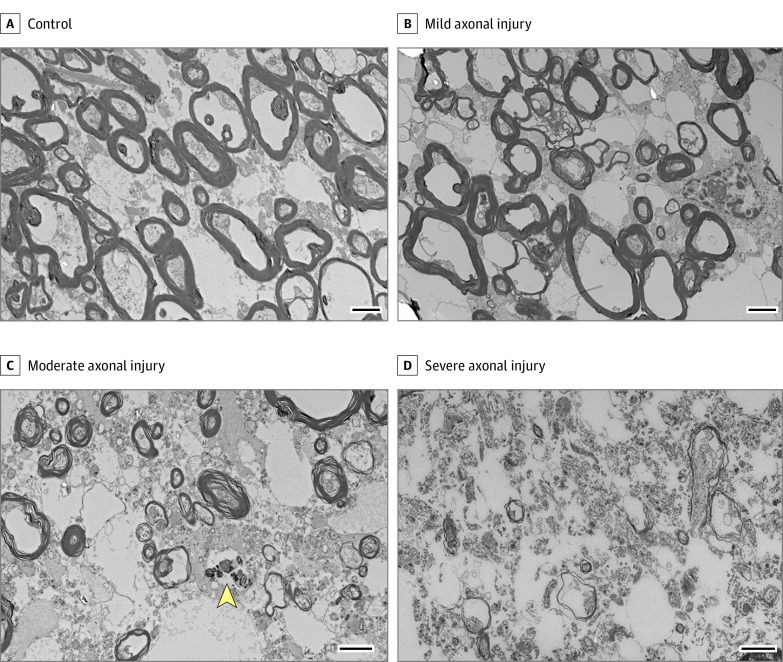
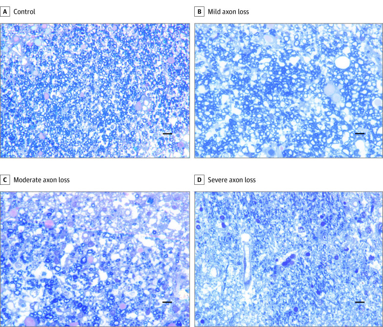
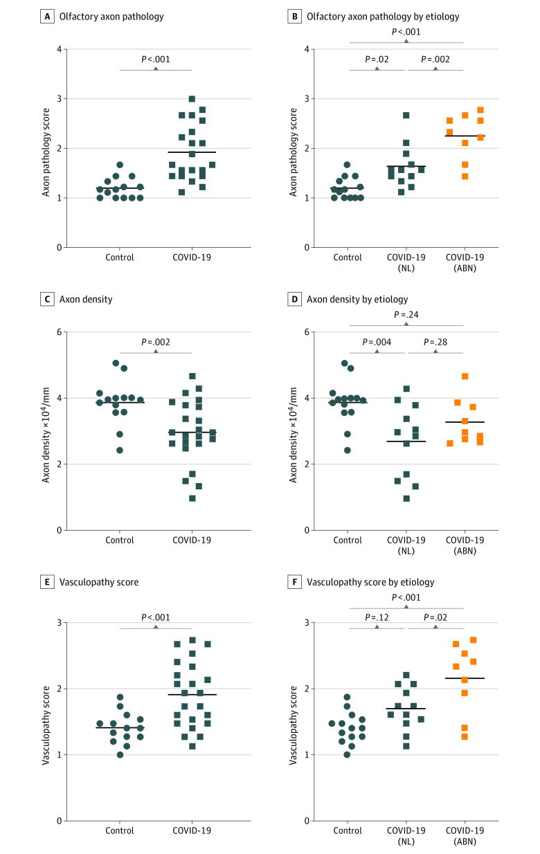
Routine histopathology was examined but did not reveal obvious abnormalities in olfactory tissue from individuals with COVID-19 (eFigure 1 in the Supplement). There was also no obvious increase in lymphocytic infiltrates, microglia, or reactive astrocytes in COVID-19 olfactory tissue (eTable 4 in the Supplement). Conversely, semithin section and ultrastructural analyses by electron microscopy demonstrated a spectrum of axon pathology ranging from mild changes to severe axonal injury and axonal losses (Figure 1, Figure 2, and Table). Axon pathology was further scored by 3 board-certified neuropathologists based on the criteria listed in eTable 2 in the Supplement. Compared with controls, individuals with COVID-19 showed significantly worse olfactory axonal damage (Figure 3 and Table). The mean (SD) axon pathology score (range, 1-3) was 1.921 (0.569) in patients with COVID-19 and 1.198 (0.208) in controls (95% CI, 0.444-1.002; P < .001). The mean (SD) axon density in lateral olfactory tract was 2.973 (0.963) × 104/mm2 in patients with COVID-19 and 3.867 (0.670) × 104/mm2 in controls (95% CI, 0.347-1.440; P = .002), indicating a 23% loss of olfactory axons. Overall, individuals with reported smell alterations had significantly more severe olfactory axon pathology than cases with intact smell (Figure 3). The mean (SD) axon pathology score was 2.26 (0.457) in patients with COVID-19 with smell alterations and 1.63 (0.426) in patients with intact smell (95% CI, 0.229-1.031; P = .002). However, there was no significant difference in axon densities between patients with COVID-19 with smell alterations (mean [SD], 3.272 [0.689] × 104/mm2) and those with intact smell (mean [SD], 2.693 [1.097] × 104/mm2; 95% CI, −0.336 to 1.496; P = .28).
Figure 1. Electron Micrographs Demonstrate a Spectrum of Olfactory Axonal Injuries in Patients With COVID-19 .
A spectrum of axonal degeneration including organelle aggregation, myelin sheath disintegration, myelin sheath vacuolization, and myelin debris can be seen in COVID-19 cases. Scale bars represent 2 μm. The arrowhead indicates myelin debris.
Figure 2. Axon Losses Ranging From Mild to Severe Are Observed in Toluidine Blue-Stained Semithin Sections of the Lateral Olfactory Tract From Patients With COVID-19 .
Scale bars represent 10 μm.
Figure 3. Olfactory Axon Pathology, Axon Losses, and Microvasculopathy Are Significantly Worse in Patients With COVID-19 Than Control Individuals.
A, C, and E, Comparisons between individuals with COVID-19 and control individuals (t test). Individuals with COVID-19 are further divided into smell normal (NL) and smell abnormal (ABN) groups for the comparisons in B, D, and F (1-way analysis of variance).
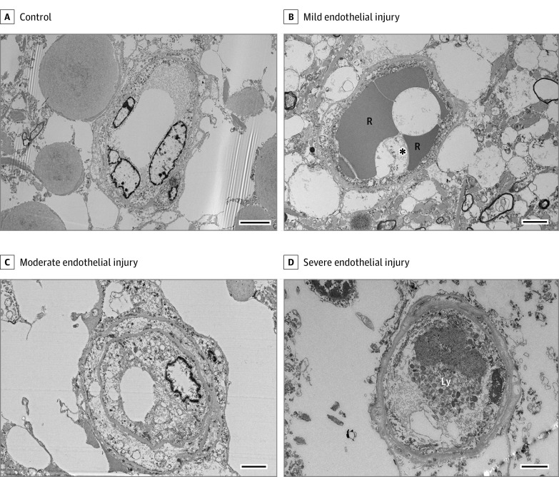
In addition to axon pathology, individuals with COVID-19 were remarkable for endothelial injury of the microvasculature, an immune-mediated process seen primarily in the heart and lung.27,28 Ultrastructural findings, including endothelial cytoplasmic swelling, vacuolization, structural damage of mitochondria, and subendothelial edema, were frequently observed in patients with COVID-19 (Figure 4). The mean (SD) microvasculopathy score (range, 1-3) was 1.907 (0.490) in patients with COVID-19 and 1.405 (0.233) in control individuals (95% CI, 0.259-0.745; P < .001). Similar to axon pathology, patients with COVID-19 with smell alterations also had a worse microvascular pathology score (mean [SD], 2.154 [0.528]) than those with intact smell (mean [SD], 1.694 [0.329]; 95% CI, 0.071-0.850; P = .02). Notably, mild microvascular injury was present in control individuals, suggesting that some degree of endothelial abnormalities may be an end-of-life change rather than a pathological condition. In comparison, moderate to severe endothelial injuries were noted particularly in patients with COVID-19 with abnormal sense of smell and therefore may be clinically significant.
Figure 4. Electron Micrographs of Olfactory Microvasculature From Patients With COVID-19 Demonstrate a Spectrum of Endothelial Injuries.
A spectrum of microvascular endothelial injury including cytoplasmic swelling, cytoplasmic vacuolization, ultrastructural abnormalities in mitochondria, and lysosome accumulation can be seen in patients with COVID-19. Note the occlusion of the capillary lumen by the swollen cytoplasm of the endothelial cells in the severe case (D). Scale bars represent 2 μm. The asterisk indicates a cytoplasmic vacuole. Ly indicates lysosome; R, red blood cell.
Of note, olfactory axon and microvascular pathology do not appear to be associated with clinical severity or timing of the infection. In addition, SARS-CoV-2 was only detected in olfactory tissue from 3 patients by droplet digital PCR or immunofluorescence (Table), suggesting that olfactory pathology was not caused by direct viral injury.
Because advancing age has been previously associated with neuropathologic changes, eg, tau deposits, multivariable linear regression was conducted to test whether the association of COVID-19 with axon and vascular pathology was independent of age. After controlling for age, COVID-19 diagnosis remained associated with increased axonal pathology score (β, 0.774; 95% CI, 0.439-1.110; P < .001), reduced axonal density (β, −0.661; 95% CI, −1.209 to −0.114; P = .02), and increased vascular pathology score (β, 0.522; 95% CI, 0.222-0.822; P = .001). Nevertheless, age was associated with decreasing axon densities independent of COVID-19 status (β, −0.028; 95% CI, −0.043 to −0.013; P < .001; eFigure 2 in the Supplement).
To further address whether age-associated tau pathology contributed to olfactory axon pathology, phospho-tau immunohistochemical stain was performed on olfactory tissue from both patients with COVID-19 and control individuals. In addition, neuropathologic evaluation for Alzheimer disease was performed on patients with COVID-19 and control individuals older than 40 years (eTable 5 in the Supplement). The COVID-19 group with smell alterations had 2 patients with low-level Alzheimer disease neuropathologic change; the COVID-19 group with intact smell had 1 patient with intermediate-level and 3 patients with low-level change; and the control group had 1 individual with intermediate-level and 2 individuals with low-level change. All 3 groups had 2 patients with Braak stage higher than II. In terms of tau deposits in olfactory tissue, the COVID-19 group with smell alterations had 1 patient with moderate neurofibrillary tangles (NFTs), and the COVID-19 group with intact smell had 2 patients with frequent and 1 patient with moderate NFTs. The frequency of NFTs in olfactory tissue roughly associated with the Braak staging. Although the control group did not have any individual with moderate or frequent NFTs in olfactory tissue, this might be because the 2 control individuals with higher Braak staging had no sufficient olfactory tissue for assessment. Overall, patients with COVID-19 did not have more severe age-related tau pathology in brain or olfactory tissue than control individuals. This finding further supported the results from the multivariable analyses that the difference in olfactory pathology between patients with COVID-19 and controls could not simply be accounted for by the age difference.
It was noteworthy that some cases in this study had a prolonged postmortem interval. While the autolytic process associated with prolonged postmortem interval may cause changes in ultrastructures of axons and vessels, patients with longer postmortem interval did not appear to have worse axon or microvascular pathology within each group (Table).
Discussion
The olfactory pathway begins with ORNs in the nasal mucosa. ORNs extend their axons to the olfactory bulb to form synapses with mitral cells and tufted cells, the second-order olfactory neurons in the olfactory bulb. These neurons then project their axons through the lateral olfactory tract into the olfactory cortex. Olfactory dysfunction can occur if any of the components in the olfactory pathway gets injured.
Olfactory dysfunction is most commonly associated with viral infection of the upper respiratory tract.29,30 The role of viral injuries in postviral olfactory dysfunction remains unclear; some studies reveal near-complete degeneration of the ORNs in nasal mucosa, whereas others show unremarkable histology.29,31 A study from Meinhardt and colleagues16 examined the nasal mucosa from patients with COVID-19 and demonstrated the capability of SARS-CoV-2 to infect ORNs. Another study from Khan and colleagues19 showed that nonneuronal sustentacular cells in the nasal mucosa were in fact the main target for SARS-CoV-2. While the pathologic effect of COVID-19 infection on nasal mucosa may be sufficient to cause olfactory dysfunction, it fails to explain the persistent anosmia experienced by some patients4,6,11,12 since cell damage in nasal mucosa is largely repairable.
To our knowledge, this cohort study was the first to examine the ultrastructural changes of olfactory bulb and olfactory tract in COVID-19 infection. Previous studies of olfactory bulb have been limited to conventional histologic examination without providing appropriate controls or information regarding patients’ smell function.23,32 Results from this study not only confirmed at an ultrastructural level the previously reported radiological abnormalities in olfactory bulb33,34 but also demonstrated how the pathologic findings were associated with smell alterations in patients with COVID-19. Consistent with findings reported by other groups,16,19 this study also did not find evidence of viral infection in olfactory bulb from most patients with COVID-19. Therefore, the axon and microvascular pathology in olfactory bulb and olfactory tract were most likely not caused by direct viral injury.
Endothelial cell injury and dysfunction is a common phenomenon in COVID-19 infection, particularly in the heart and lung.27,28 It is thought to be caused by excessive cytokine release from immune cells, respiratory epithelial cells, and alveolar epithelial cells and has been shown to correlate with COVID-19 severity.28 However, this study did not find a strong association between olfactory endothelial injury and disease severity, suggesting that local inflammation in the upper respiratory tract may be sufficient to cause endothelial and axonal damage in the olfactory pathway. Schwabenland and colleagues35 performed deep spatial profiling and found a mild increase in lymphocytes and microglia in olfactory bulb and brain stem from patients with COVID-19.Although we did not see an obvious overall increase in inflammatory cells in our cohort, it remained possible that focal or perivascular infiltrates may have a role in axonal or microvascular injuries associated with COVID-19 infection.
Limitations
This study did not examine the pathologic association of COVID-19 with nasal mucosa, which contains primary olfactory neurons and their supporting cells. Therefore, how pathologic changes in nasal mucosa may contribute to smell alterations in COVID-19 infection could not be assessed in this cohort.
In this study, information regarding smell function was obtained by subjective self-report or reporting from patients’ family members. Objective olfactory function assessment would be considered medically unnecessary for patients with serious illness and was impossible for patients receiving mechanical ventilation. While subjective self-report may not accurately reflect olfactory function, the issue appears to be underreporting rather than overreporting of olfactory dysfunction.36
Conclusions
The results of this postmortem cohort study demonstrated that COVID-19 infection could cause axon injuries and microvasculopathy in olfactory tissue. Overall, patients with smell alterations had more severe olfactory pathology than those with intact smell. While most individuals in our study demonstrated mild to moderate findings, the severity of pathology in some COVID-19 cases indicates that the olfactory system damage and dysfunction can be permanent.
eTable 1. Demographics, medical history and significant autopsy findings
eTable 2. Scoring criteria for axon and microvascular pathology
eTable 3. Interobserver reliability in axon and microvascular pathology scoring
eTable 4. No increase in neuroinflammation in olfactory tissue from COVID-19 patients
eTable 5. No significant tau deposits in olfactory or brain tissue from COVID-19 patients
eFigure 1. COVID-19 olfactory bulb and olfactory tract are unremarkable on routine histological examination
eFigure 2. Multiple linear regression analysis assessing the effect of age on the following olfactory pathologic features: (A) Axon pathology (B) Axon density, and (C) Microvascular pathology
References
- 1.Agyeman AA, Chin KL, Landersdorfer CB, Liew D, Ofori-Asenso R. Smell and taste dysfunction in patients with COVID-19: a systematic review and meta-analysis. Mayo Clin Proc. 2020;95(8):1621-1631. doi: 10.1016/j.mayocp.2020.05.030 [DOI] [PMC free article] [PubMed] [Google Scholar]
- 2.Carignan A, Valiquette L, Grenier C, et al. Anosmia and dysgeusia associated with SARS-CoV-2 infection: an age-matched case-control study. CMAJ. 2020;192(26):E702-E707. doi: 10.1503/cmaj.200869 [DOI] [PMC free article] [PubMed] [Google Scholar]
- 3.Giacomelli A, Pezzati L, Conti F, et al. Self-reported olfactory and taste disorders in patients with severe acute respiratory coronavirus 2 infection: a cross-sectional study. Clin Infect Dis. 2020;71(15):889-890. doi: 10.1093/cid/ciaa330 [DOI] [PMC free article] [PubMed] [Google Scholar]
- 4.Moein ST, Hashemian SM, Tabarsi P, Doty RL. Prevalence and reversibility of smell dysfunction measured psychophysically in a cohort of COVID-19 patients. Int Forum Allergy Rhinol. 2020;10(10):1127-1135. doi: 10.1002/alr.22680 [DOI] [PMC free article] [PubMed] [Google Scholar]
- 5.Moein ST, Hashemian SM, Mansourafshar B, Khorram-Tousi A, Tabarsi P, Doty RL. Smell dysfunction: a biomarker for COVID-19. Int Forum Allergy Rhinol. 2020;10(8):944-950. doi: 10.1002/alr.22587 [DOI] [PMC free article] [PubMed] [Google Scholar]
- 6.Lechien JR, Chiesa-Estomba CM, Beckers E, et al. Prevalence and 6-month recovery of olfactory dysfunction: a multicentre study of 1363 COVID-19 patients. J Intern Med. 2021;290(2):451-461. doi: 10.1111/joim.13209 [DOI] [PubMed] [Google Scholar]
- 7.Zahra SA, Iddawela S, Pillai K, Choudhury RY, Harky A. Can symptoms of anosmia and dysgeusia be diagnostic for COVID-19? Brain Behav. 2020;10(11):e01839. doi: 10.1002/brb3.1839 [DOI] [PMC free article] [PubMed] [Google Scholar]
- 8.Cho RHW, To ZWH, Yeung ZWC, et al. COVID-19 viral load in the severity of and recovery from olfactory and gustatory dysfunction. Laryngoscope. 2020;130(11):2680-2685. doi: 10.1002/lary.29056 [DOI] [PMC free article] [PubMed] [Google Scholar]
- 9.Sayin İ, Yaşar KK, Yazici ZM. Taste and smell impairment in COVID-19: an AAO-HNS anosmia reporting tool-based comparative study. Otolaryngol Head Neck Surg. 2020;163(3):473-479. doi: 10.1177/0194599820931820 [DOI] [PMC free article] [PubMed] [Google Scholar]
- 10.Xydakis MS, Dehgani-Mobaraki P, Holbrook EH, et al. Smell and taste dysfunction in patients with COVID-19. Lancet Infect Dis. 2020;20(9):1015-1016. doi: 10.1016/S1473-3099(20)30293-0 [DOI] [PMC free article] [PubMed] [Google Scholar]
- 11.Hopkins C, Surda P, Whitehead E, Kumar BN. Early recovery following new onset anosmia during the COVID-19 pandemic: an observational cohort study. J Otolaryngol Head Neck Surg. 2020;49(1):26. doi: 10.1186/s40463-020-00423-8 [DOI] [PMC free article] [PubMed] [Google Scholar]
- 12.Renaud M, Thibault C, Le Normand F, et al. Clinical outcomes for patients with anosmia 1 year after COVID-19 diagnosis. JAMA Netw Open. 2021;4(6):e2115352-e2115352. doi: 10.1001/jamanetworkopen.2021.15352 [DOI] [PMC free article] [PubMed] [Google Scholar]
- 13.Aragão MFVV, Leal MC, Cartaxo Filho OQ, Fonseca TM, Valença MM. Anosmia in COVID-19 associated with injury to the olfactory bulbs evident on MRI. AJNR Am J Neuroradiol. 2020;41(9):1703-1706. doi: 10.3174/ajnr.A6675 [DOI] [PMC free article] [PubMed] [Google Scholar]
- 14.Eliezer M, Hamel AL, Houdart E, et al. Loss of smell in patients with COVID-19: MRI data reveal a transient edema of the olfactory clefts. Neurology. 2020;95(23):e3145-e3152. doi: 10.1212/WNL.0000000000010806 [DOI] [PubMed] [Google Scholar]
- 15.Laurendon T, Radulesco T, Mugnier J, et al. Bilateral transient olfactory bulb edema during COVID-19-related anosmia. Neurology. 2020;95(5):224-225. doi: 10.1212/WNL.0000000000009850 [DOI] [PubMed] [Google Scholar]
- 16.Meinhardt J, Radke J, Dittmayer C, et al. Olfactory transmucosal SARS-CoV-2 invasion as a port of central nervous system entry in individuals with COVID-19. Nat Neurosci. 2021;24(2):168-175. doi: 10.1038/s41593-020-00758-5 [DOI] [PubMed] [Google Scholar]
- 17.Bryche B, St Albin A, Murri S, et al. Massive transient damage of the olfactory epithelium associated with infection of sustentacular cells by SARS-CoV-2 in golden Syrian hamsters. Brain Behav Immun. 2020;89:579-586. doi: 10.1016/j.bbi.2020.06.032 [DOI] [PMC free article] [PubMed] [Google Scholar]
- 18.Sia SF, Yan LM, Chin AWH, et al. Pathogenesis and transmission of SARS-CoV-2 in golden hamsters. Nature. 2020;583(7818):834-838. doi: 10.1038/s41586-020-2342-5 [DOI] [PMC free article] [PubMed] [Google Scholar]
- 19.Khan M, Yoo SJ, Clijsters M, et al. Visualizing in deceased COVID-19 patients how SARS-CoV-2 attacks the respiratory and olfactory mucosae but spares the olfactory bulb. Cell. 2021;184(24):5932-5949.e15. doi: 10.1016/j.cell.2021.10.027 [DOI] [PMC free article] [PubMed] [Google Scholar]
- 20.Bilinska K, Jakubowska P, Von Bartheld CS, Butowt R. Expression of the SARS-CoV-2 entry proteins, ACE2 and TMPRSS2, in cells of the olfactory epithelium: identification of cell types and trends with age. ACS Chem Neurosci. 2020;11(11):1555-1562. doi: 10.1021/acschemneuro.0c00210 [DOI] [PMC free article] [PubMed] [Google Scholar]
- 21.Brann DH, Tsukahara T, Weinreb C, et al. Non-neuronal expression of SARS-CoV-2 entry genes in the olfactory system suggests mechanisms underlying COVID-19-associated anosmia. Sci Adv. 2020;6(31):eabc5801. doi: 10.1126/sciadv.abc5801 [DOI] [PMC free article] [PubMed] [Google Scholar]
- 22.Bilinska K, Butowt R. Anosmia in COVID-19: a bumpy road to establishing a cellular mechanism. ACS Chem Neurosci. 2020;11(15):2152-2155. doi: 10.1021/acschemneuro.0c00406 [DOI] [PMC free article] [PubMed] [Google Scholar]
- 23.Solomon IH, Normandin E, Bhattacharyya S, et al. Neuropathological features of Covid-19. N Engl J Med. 2020;383(10):989-992. doi: 10.1056/NEJMc2019373 [DOI] [PMC free article] [PubMed] [Google Scholar]
- 24.Mukerji SS, Solomon IH. What can we learn from brain autopsies in COVID-19? Neurosci Lett. 2021;742:135528. doi: 10.1016/j.neulet.2020.135528 [DOI] [PMC free article] [PubMed] [Google Scholar]
- 25.Landis JR, Koch GG. An application of hierarchical kappa-type statistics in the assessment of majority agreement among multiple observers. Biometrics. 1977;33(2):363-374. doi: 10.2307/2529786 [DOI] [PubMed] [Google Scholar]
- 26.Engelmann S, Ruewe M, Geis S, et al. Rapid and precise semi-automatic axon quantification in human peripheral nerves. Sci Rep. 2020;10(1):1935. doi: 10.1038/s41598-020-58917-4 [DOI] [PMC free article] [PubMed] [Google Scholar]
- 27.Varga Z, Flammer AJ, Steiger P, et al. Endothelial cell infection and endotheliitis in COVID-19. Lancet. 2020;395(10234):1417-1418. doi: 10.1016/S0140-6736(20)30937-5 [DOI] [PMC free article] [PubMed] [Google Scholar]
- 28.Jin Y, Ji W, Yang H, Chen S, Zhang W, Duan G. Endothelial activation and dysfunction in COVID-19: from basic mechanisms to potential therapeutic approaches. Signal Transduct Target Ther. 2020;5(1):293. doi: 10.1038/s41392-020-00454-7 [DOI] [PMC free article] [PubMed] [Google Scholar]
- 29.Jafek BW, Murrow B, Michaels R, Restrepo D, Linschoten M. Biopsies of human olfactory epithelium. Chem Senses. 2002;27(7):623-628. doi: 10.1093/chemse/27.7.623 [DOI] [PubMed] [Google Scholar]
- 30.Temmel AFP, Quint C, Schickinger-Fischer B, Klimek L, Stoller E, Hummel T. Characteristics of olfactory disorders in relation to major causes of olfactory loss. Arch Otolaryngol Head Neck Surg. 2002;128(6):635-641. doi: 10.1001/archotol.128.6.635 [DOI] [PubMed] [Google Scholar]
- 31.Moran DT, Jafek BW, Eller PM, Rowley JC III. Ultrastructural histopathology of human olfactory dysfunction. Microsc Res Tech. 1992;23(2):103-110. doi: 10.1002/jemt.1070230202 [DOI] [PubMed] [Google Scholar]
- 32.Lee M-H, Perl DP, Nair G, et al. Microvascular injury in the brains of patients with Covid-19. N Engl J Med. 2021;384(5):481-483. doi: 10.1056/NEJMc2033369 [DOI] [PMC free article] [PubMed] [Google Scholar]
- 33.Chiu A, Fischbein N, Wintermark M, Zaharchuk G, Yun PT, Zeineh M. COVID-19-induced anosmia associated with olfactory bulb atrophy. Neuroradiology. 2021;63(1):147-148. doi: 10.1007/s00234-020-02554-1 [DOI] [PMC free article] [PubMed] [Google Scholar]
- 34.Kandemirli SG, Altundag A, Yildirim D, Tekcan Sanli DE, Saatci O. Olfactory bulb MRI and paranasal sinus CT findings in persistent COVID-19 anosmia. Acad Radiol. 2021;28(1):28-35. doi: 10.1016/j.acra.2020.10.006 [DOI] [PMC free article] [PubMed] [Google Scholar]
- 35.Schwabenland M, Salié H, Tanevski J, et al. Deep spatial profiling of human COVID-19 brains reveals neuroinflammation with distinct microanatomical microglia-T-cell interactions. Immunity. 2021;54(7):1594-1610.e11. doi: 10.1016/j.immuni.2021.06.002 [DOI] [PMC free article] [PubMed] [Google Scholar]
- 36.Mazzatenta A, Neri G, D’Ardes D, et al. Smell and taste in severe CoViD-19: self-reported vs. testing. Front Med (Lausanne). 2020;7:589409. doi: 10.3389/fmed.2020.589409 [DOI] [PMC free article] [PubMed] [Google Scholar]
Associated Data
This section collects any data citations, data availability statements, or supplementary materials included in this article.
Supplementary Materials
eTable 1. Demographics, medical history and significant autopsy findings
eTable 2. Scoring criteria for axon and microvascular pathology
eTable 3. Interobserver reliability in axon and microvascular pathology scoring
eTable 4. No increase in neuroinflammation in olfactory tissue from COVID-19 patients
eTable 5. No significant tau deposits in olfactory or brain tissue from COVID-19 patients
eFigure 1. COVID-19 olfactory bulb and olfactory tract are unremarkable on routine histological examination
eFigure 2. Multiple linear regression analysis assessing the effect of age on the following olfactory pathologic features: (A) Axon pathology (B) Axon density, and (C) Microvascular pathology


