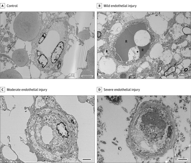
Figure 4. Electron Micrographs of Olfactory Microvasculature From Patients With COVID-19 Demonstrate a Spectrum of Endothelial Injuries.
A spectrum of microvascular endothelial injury including cytoplasmic swelling, cytoplasmic vacuolization, ultrastructural abnormalities in mitochondria, and lysosome accumulation can be seen in patients with COVID-19. Note the occlusion of the capillary lumen by the swollen cytoplasm of the endothelial cells in the severe case (D). Scale bars represent 2 μm. The asterisk indicates a cytoplasmic vacuole. Ly indicates lysosome; R, red blood cell.

