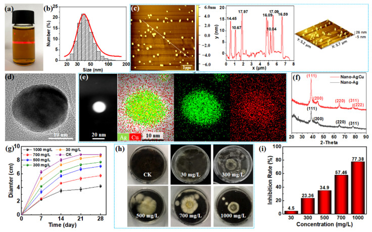
Figure 2.
Characterization of nano-AgCu and its inhibition effect against Aspergillus niger on the PDA plates. (a) The aqueous nano-AgCu suspension passed through by a laser light; (b) the size distribution histogram of nano-AgCu particles; (c) AFM images of nano-AgCu particles; (d,e) HRTEM-EDX images of nano-AgCu particle; (f) XRD patterns of nano-AgCu and corresponding nano-Ag particles; (g) variations of growth diameter of fungus on the PDA plate with growing time at different nano-AgCu concentrations; (h) digital photos of fungal growth on the nano-AgCu-treated PDA plate at different concentrations after 28 days; (i) inhibition rate of nano-AgCu against fungal growth on the PDA plate at different concentrations.

