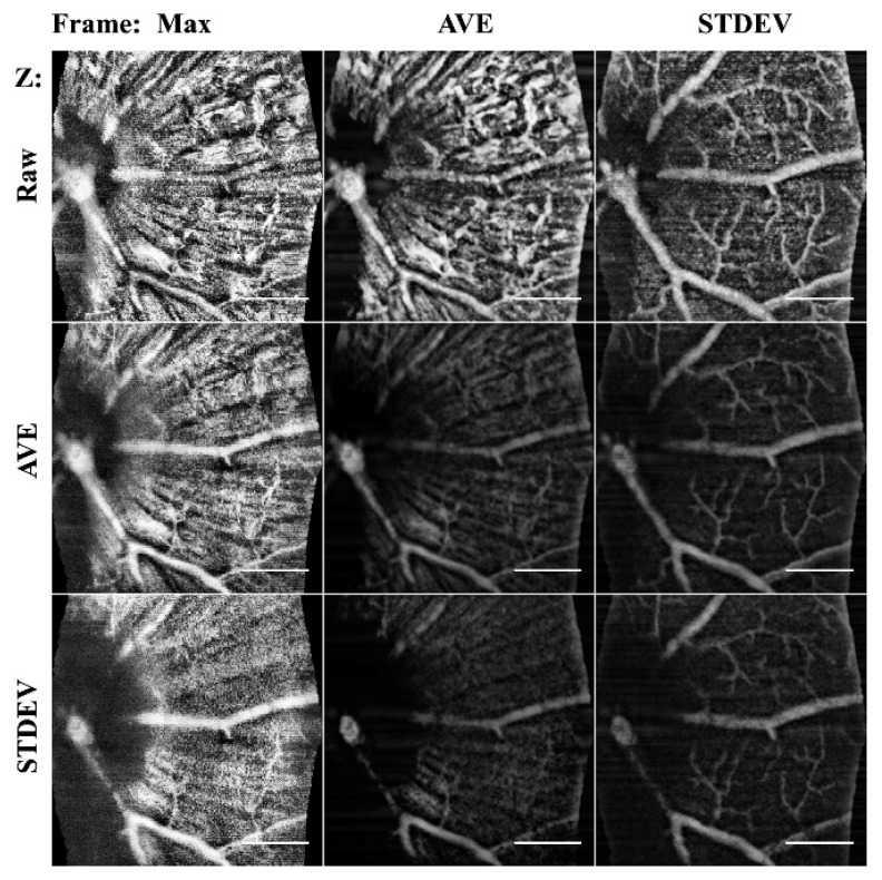
Figure 4.
Comparison of frame averaging methods. Columns are the frame averaging method used to project the 60 μm stack, and the rows are the input b-scan averaging method used. By altering the combinations, the specificity to detect different the morphology is selectable. STDEV frame averaging improved vessels, where mean AVE enhanced NFL. MAX intensity projections only captured the ILM. Scale bar 200 μm.

