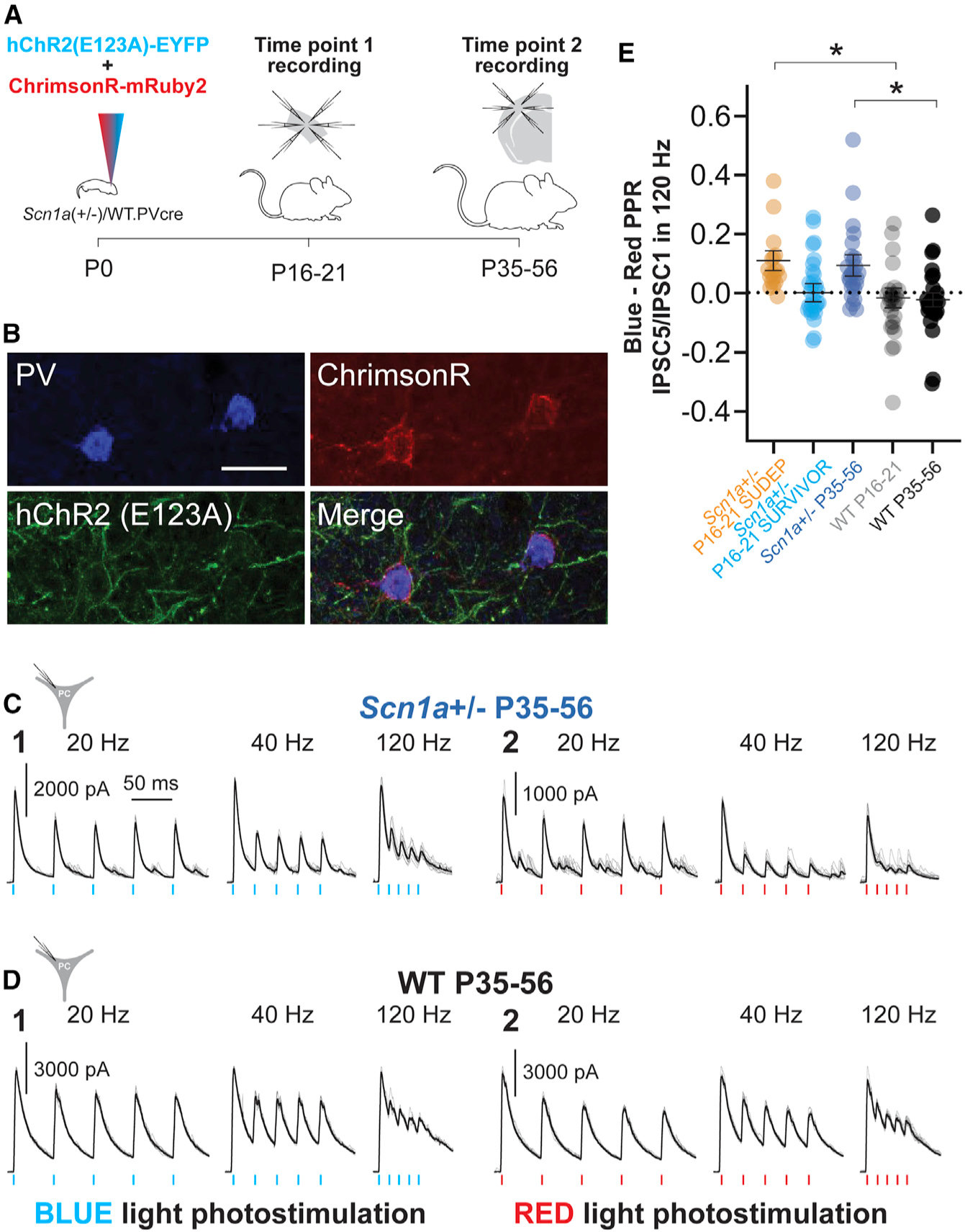
Figure 7. A dual optogenetics strategy demonstrates impaired PV interneuron spike propagation in Scn1a+/− mice.

(A) Experimental timeline showing viral injection of both AAV9.DIO.ChrimsonR.mRuby2 and AAV.DIO.hChR2.eYFP into PV-Cre mice at P0 (left) and time points 1 (mini-slice; center) and 2 (acute brain slice; right) recordings.
(B) Representative confocal images showing expression of PV (Alexa Fluor 405; blue), hChR2.eYFP (green), and ChrimsonR.mRuby2 (red). Scale bar, 20 μm.
(C and D) Representative data for evoked oIPSCs recorded in principal cells in response to repetitive photostimulation with ChR2 (C1 and D1) or ChrimsonR (C2 and D2) in Scn1a+/− P35–P56 mice
(C) and WT P35–P56 mice (D).
(E) Summary data for the index of oIPSCn/oIPSC1 in response to blue-light photostimulation minus oIPSCn/oIPSC1 in response to red-light photostimulation (blue-red index). *p<0.05 via mixed-effects modeling.
See Table S3.
