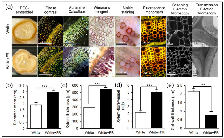
Figure 3.
Qualitative and quantitative analyses of the structural and ultrastructural changes, as well as of the cell wall components of the stems of tobacco plants grown under White and White + FR conditions. Samples were fixed in Karnovsky’s solution, embedded in PEG 6000 and analysed using different staining techniques followed by fluorescence and electron microscopy. (a) From left to right: stem sections embedded in PEG 6000; stained with toluidine blue and analysed under light microscopy with phase contrast; blocks in methyl methacrylate stained with Auramine O and Calcofluor Bright White 28 and analysed using epifluorescence microscopy; lignified cells stained with Wiesner’s reagent and analysed under light microscopy; Mäule reagent showing lignin monomers under light microscopy; staining with astra blue and safranin-fuchsin followed by fluorescence microscopy indicate changes in the monomeric composition of lignin; superficial changes in the texture and structure of the cell wall revealed via scanning electron microscopy (SEM) and changes in the organisation and arrangement of cellulose microfibrils revealed via transmission electron microscopy (TEM). (b) Stem diameter of the medial region (cm). (c) Xylem thickness (µm). (d) Xylem fibre-like-cells/vessel ratio. (e) Cell wall thickness (µm). Asterisks (***) indicate statistical differences between distinct treatments using the Student’s t-test (p < 0.001); (n = 10 ± SE).

