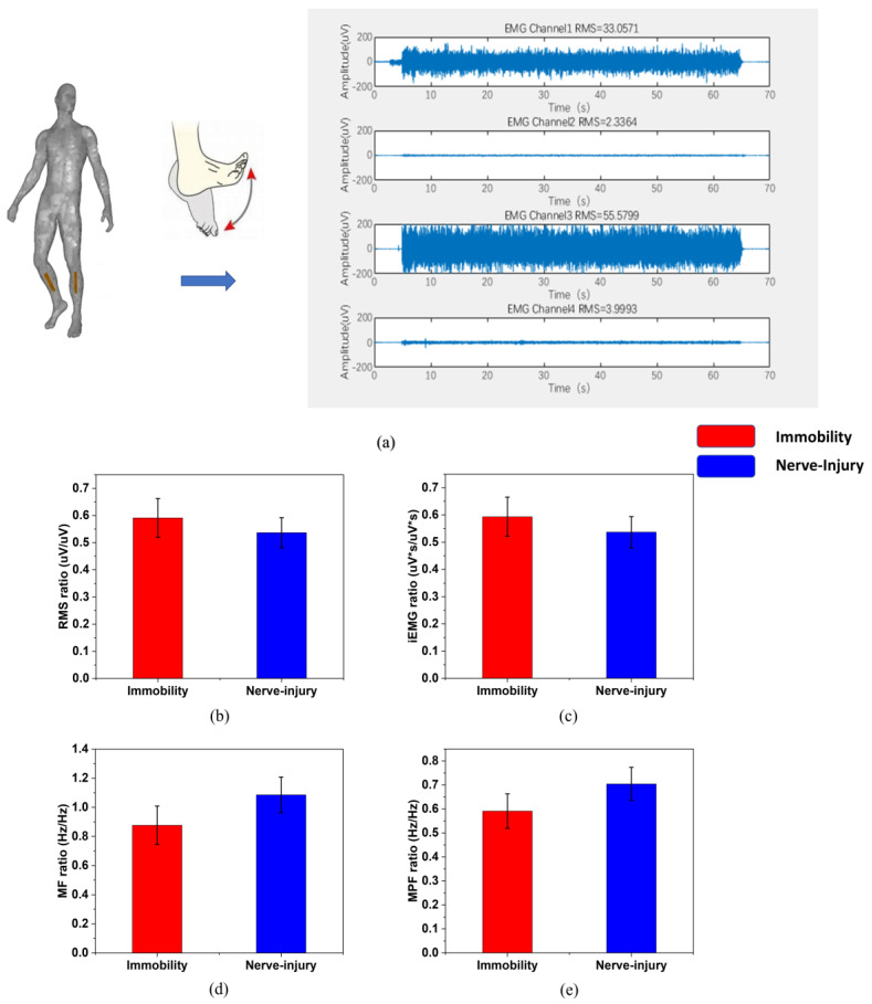
Figure 5.
(a) The protocol of the moves and the data acquisition on the affected and unaffected sides. (The four channels are: tibialis anterior muscle of the affected limb, gastrocnemius muscle of the affected limb, tibialis anterior muscle of the unaffected limb, gastrocnemius muscle of the unaffected limb). The sEMG features are gathered every 0.5 s with an overlap of 1/8 after choosing the stable 30 s-period or 40 s-period from the 60 s-move. The distribution of (b) RMS, (c) iEMG, (d) MF, (e) MPF between nerve injury and limb immobilization patients after bone fracture.

