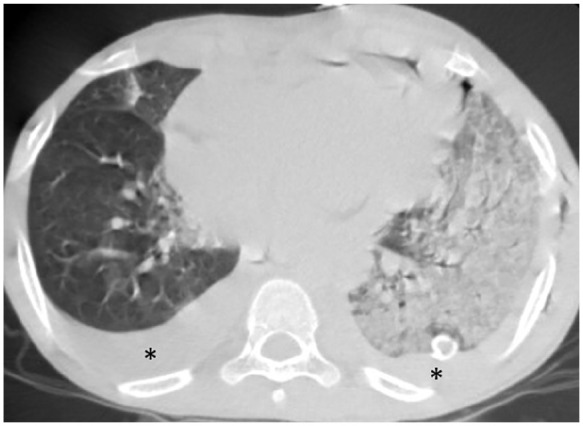
Figure 2.

12-year-old boy with known case of acute lymphoblastic leukemia, presented with fever for 4 days, conjunctivitis, maculopapular rash, hypotension and cardiogenic shock he was ventilated due respiratory distress, his COVID status was PCR swab positive , COVID IgM negative COVID IgG positive , ; axial chest CT shows extensive consolidation implicating the left lung (CT severity score= 13). Note the associated pleural effusion on both sides (asterisk). The patient was on ventilatory support.
