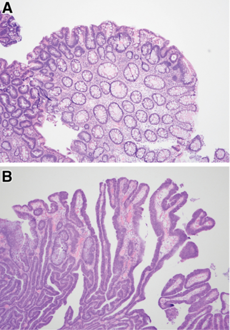
Figure 3.
Histologic examples of conventional dysplasia. (A) Sporadic adenoma demonstrating dysplastic crypts at the surface of the polyp (“top-down”). (B) Colitis-associated dysplastic lesion demonstrating dysplastic cells occupying the full height of the crypts. Both are examples of low-grade dysplasia. Courtesy of Noam Harpaz, MD, PhD.

