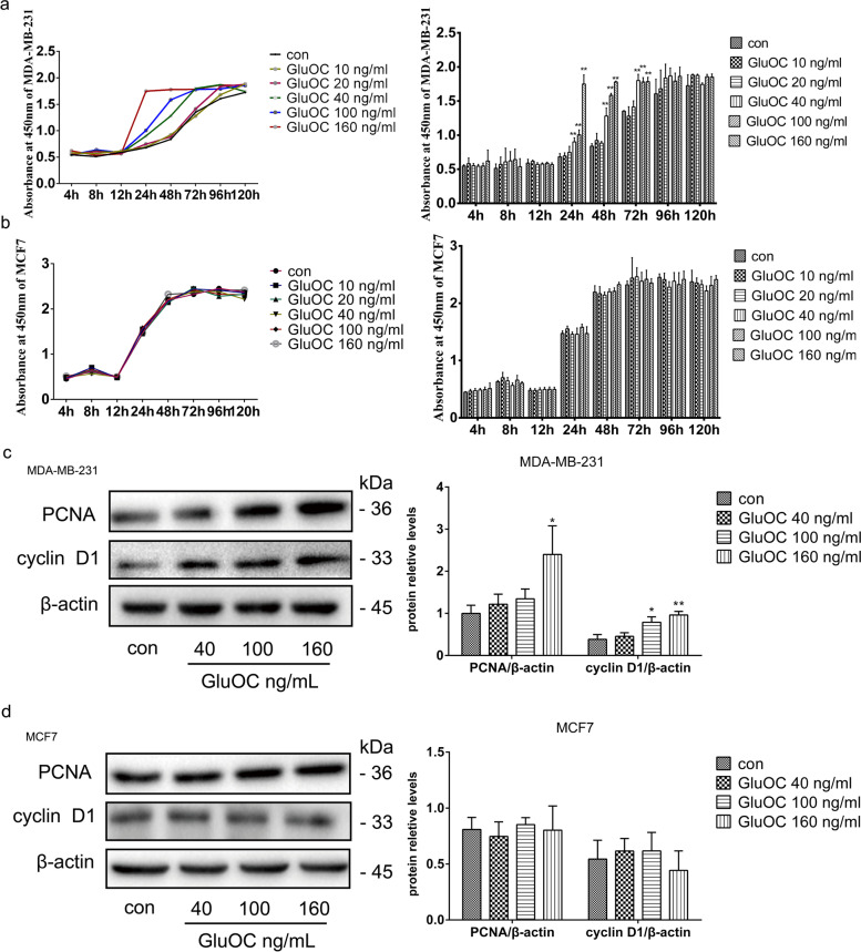
Fig. 1.
GluOC promotes MDA-MB-231 cell proliferation. a Growth curves of MDA-MB-231 cells treated with different doses of GluOC (10, 20, 40, 100, and 160 ng/ml) for different times (8, 12, 24, 48, 72, 96, and 120 h). b Growth curves of MCF7 cells treated with various doses of GluOC (10, 20, 40, 100 and 160 ng/ml) for different times (8, 12, 24, 48, 72, 96, and 120 h). c The protein levels of PCNA and cyclin D1 in MDA-MB-231 cells before and after GluOC stimulation were determined by western blotting and quantified densitometrically with ImageJ software. β-Actin was used as the control. d The protein levels of PCNA and cyclin D1 in MCF7 cells before and after GluOC stimulation were detected by western blotting. The results represent at least three independent experiments, and the data are presented as the mean ± SD (n = 3). *P < 0.05 and **P < 0.01, vs. the control group (con = control)

