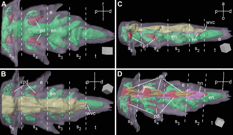
Fig. 2.
Three-dimensional reconstruction of the arm tip of A. kochii. The following anatomical structures are shown: epidermis (semitransparent violet), ectoneural part of the nervous system (green), hyponeural part of the nervous system (magenta), water-vascular system (red), arm coelom (yellow), and intervertebral muscles (brown). (a) Oral view. (b) Aboral view. (c) Side view. (d) Oblique side view, arm coelom not shown. The edge length of the scale cube (grey) is 25 m. a—aboral; c—arm coelom (somatocoel); d—distal; e—epidermis; en—ectoneural system; hn—hyponeural system; l—left; m—intervertebral muscles; o—oral; p—proximal; pd—hydrocoelic lining of the podia (tube feet); r—right; s—s—subterminal arm segments 2 thru 5; t—terminal segment; wvc—hydrocoelic water-vascular canal. The 3D video animation of this anatomical model is available in Additional file 2. The original Blender model used to produce this figure and the video is available in Zenodo (https://doi.org/10.5281/zenodo.5762494)

