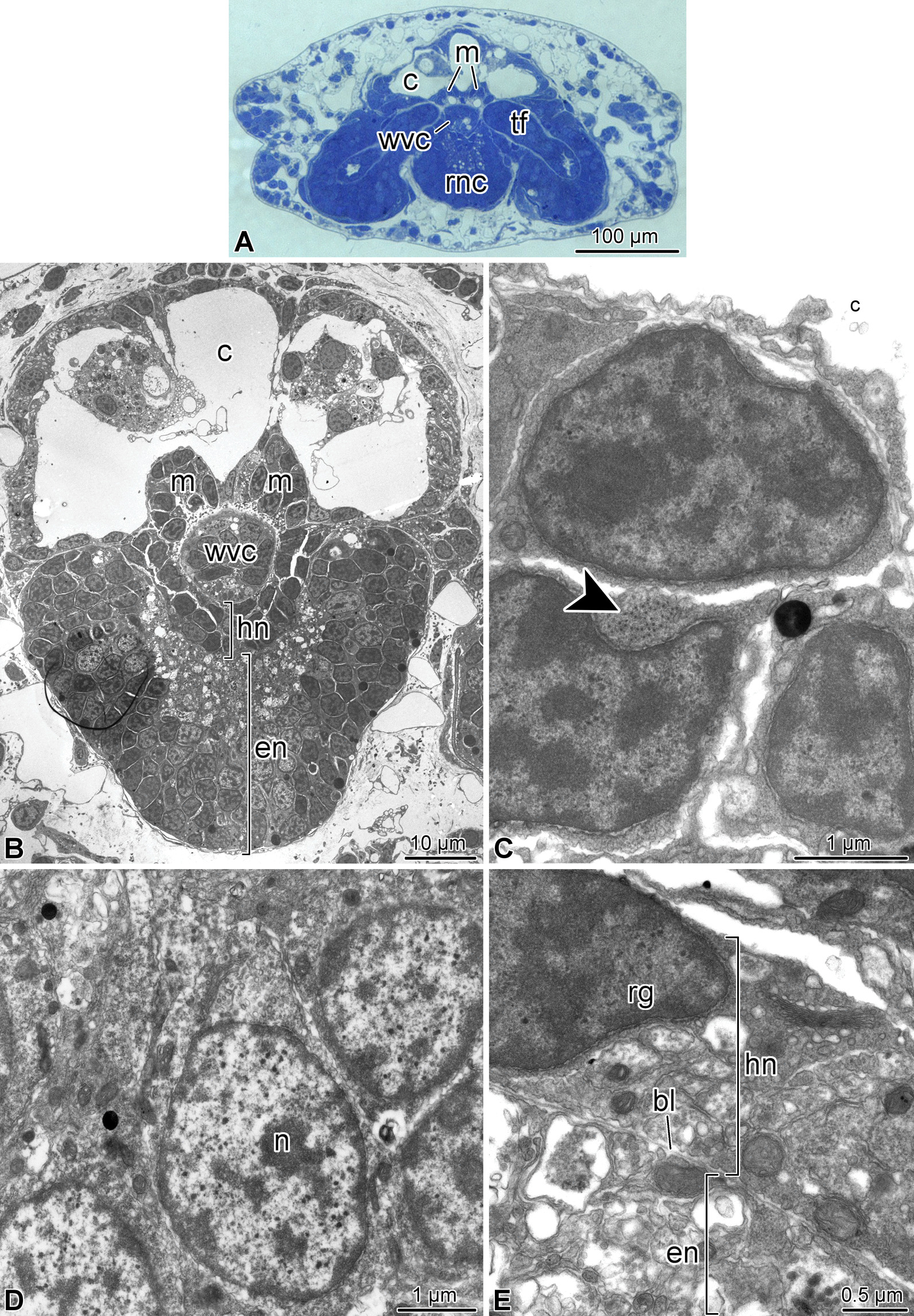
Fig. 3.

Organization of the fourth arm segment of A. kochii. Cross sections. (a) Low magnification view. Toluidine blue. (b–e) Transmission electron microscopy. (b) Low magnification transmission electron micrograph of the radial nerve cord, water-vascular canal, arm coelom, and intervertebral muscles. (c) High magnification view of the intervertebral muscle. Arrowhead shows the cytoskeletal filaments of the contractile apparatus. (d) Neuronal perikaryon in the ectoneural neuroepithelium. (e) Hyponeural part of the radial nerve cord. bl—basal lamina separating the ectoneural and hyponeural neuroepithelia; c—arm coelom; en—ectoneural neuroepithelium of the radial nerve cord; hn—hyponeural neuroepithelium of the radial nerve cord; m—intervertebral muscle; n—neuronal perikaryon; rg—radial glial cell; rnc—radial nerve cord; tf—tube foot; wvc—water-vascular canal
