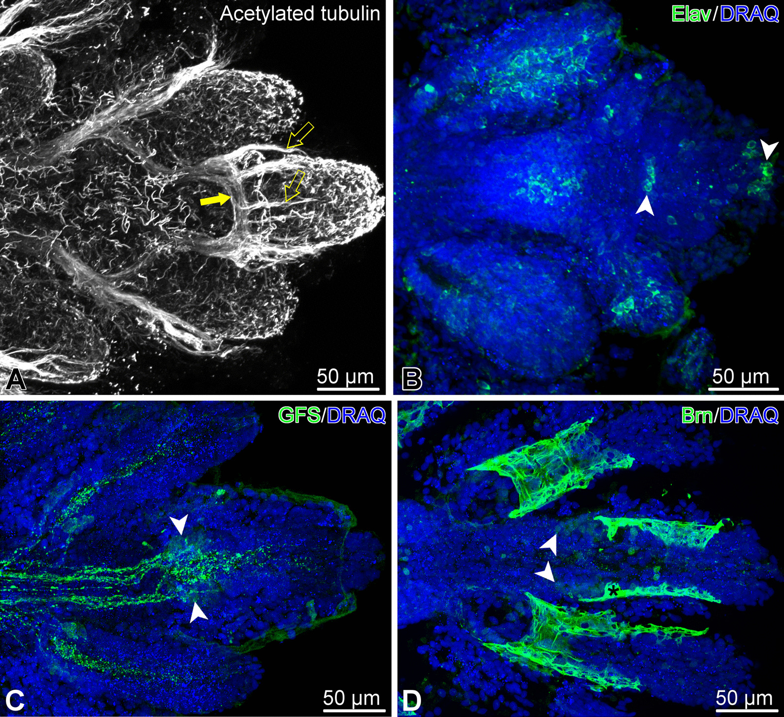
Fig. 7.

Organization of the nervous system in the arm tip of O. brevispinum. Maximum intensity Z-projections of confocal image stacks. (A) Acetylated tubulin. Filled arrow shows the terminal aboral loop formed by the hyponeural part of the radial nerve cord, which gives off a number of short tracts (open arrows) running towards the tip. (B) Elav-positive neurons. (C) GFSKLYFamide (GFS)-positive neuronal elements. (D) Expression of the transcription factor Brn1/2/4. Note that this particular antibody, besides specifically binding to the antigen in the nucleus, also non-specifically bind to the cuticle that covers the surface of the epidermis (asterisk)White arrowheads in B–D show clusters of immunopositive neuronal cell bodies
