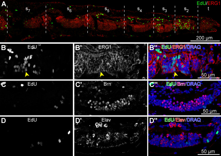
Fig. 9.
Distribution of dividing cells across the tissues and cell types in the arm tip segments O. brevispinum. The animals were pulsed with EdU for 4 h and processed for immunocytochemical staining. All micrographs are sagittal cryosections with the distal end to the right. (a) Low-magnification view of a sagittal section through the arm tip showing seven segments. (b–d”) Detailed views of the radial nerve cord in segment 2. (b–b”) Dual labeling with the EdU click reaction and ERG1 antibodies. The latter labels radial glial cells in the echinoderm central nervous system. Arrowhead points to a representative dual-labeled cell. c–d” Dual labeling with the EdU click reaction and neuronal markers Brn1/2/4 (c–c”) and Elav (d–d”). t—terminal segment; s–s—arm segments 2 thru 7, as counted from the distal tip of the arm

