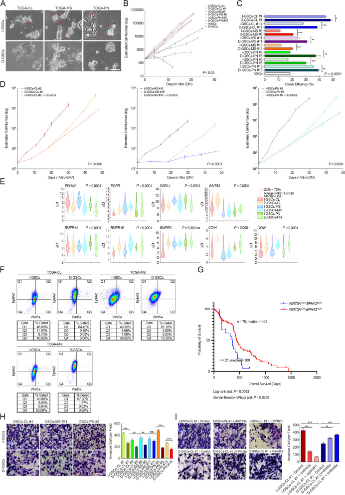
Fig. 1.
I-GSCs can be isolated from GBM surgery specimens in the absence of mitogenic stimulation. A. GFs-independent (I-GSCs) (top) and dependent GSCs (D-GSCs) (bottom) can be either isolated from the very same patient’s tissue across subtypes, with the former exhibiting many adhesion-related protrusions (arrowheads) and the latter typical rounded morphology. B-C. Significant differences in the expansion rate (B) and self-renewal potential (C) between I- and D-GSCs across subtypes, with the former comprising slower-dividing GSCs with a lower clonogenicity. *P < 0.05 I-GSCs vs. D-GSCs, hierarchical linear model for repeated measurements and ***P < 0.001, **P < 0.01, *P < 0.05 and P < 0.0001 I-GSCs vs. D-GSCs, one-way Student’s t-test in B and C, respectively. Lines I-GSCs and D-GSCs #1 (TCGA-CL GSCs, red), #6 (TCGA-MS, blue) and #15 (TCGA-PN, green) are shown as representative examples in B. Data are mean ± SD (B) and mean ± SEM (C) (n = 3). D. When exposed to mitogens, I-GSCs’ proliferation closely mirrors that one of D-GSCs, regardless of subtype (TCGA-CL GSCs, right; TCGA-MS GSCs, middle; TCGA-PN GSCs, right). ***P < 0.001 I-GSCs vs. D-GSCs, hierarchical linear model for repeated measurements. Data are mean ± SD (n = 3). E. Violin plot displaying the different enrichment of genes associated to stemness, differentiation and invasion in I-GSCs vs. D-GSCs across subtypes, as detected by qPCR. P-values are from Kruskal-Wallis test. F. Dot plots showing flow cytometric analysis confirming the enrichment of Wnt5a in I-GSCs across subtypes when compared to D-GSCs, shown to upregulate EphA2. Lines I-GSCs and D-GSCs #1 (TCGA-CL GSCs), #5 (TCGA-PN GSCs) and #11 (TCGA-MS GSCs) are shown as representative examples (n = 3). G. High level of WNT5A combined with low EPHA2 expression is associated with lower GBM patients’ survival according to TCGA public dataset (P = 0.0063 and P = 0.0259; n = 91, Log-rank and Gehan-Breslow-Wilcoxon test), as depicted by Kaplan-Meier plots. H-I. In vitro migration assay showing that, irrespective of the molecular subtype, I-GSCs migrate and invade more efficiently than their D-GSCs counterpart (H). I Blockade of Wnt5a signaling by Wnt5a-endogenous antagonists (rhWnt3a; middle and rhSFRP1; right) lessens I-GSCs’ exacerbated invasiveness (top), whereas enhancement of Wnt5a expression in D-GSCs by stable lentiviral-mediated overexpression (LV-Wnt5a; middle) or by exposure to rhWnt5a (right) elicits cell migration (bottom). Bars in A, H-I, 100um, 50um. Quantification in H-I is shown as mean ± SEM. ***P < 0.001, **P < 0.01, ns not significant, one-way Student’s t-test and ANOVA Tukey’s multiple comparison test

