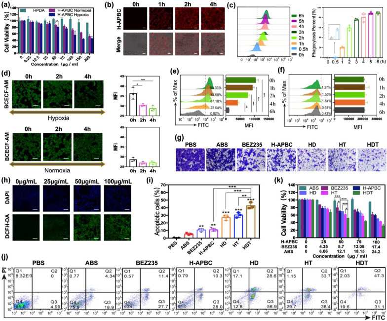
Fig. 3.
In vitro experiments with H-APBC. a Viability of 4T1 cells treated with HPDA and H-APBC without laser irradiation under the conditions of normoxic or hypoxic. (n = 3). CLSM images (b) and flow cytometric assay (c) [the average fluorescence intensity (left) and the percentage of cells taking up NPs (right)] of 4T1 cells treated with H-APBC at different time points under hypoxia. (n = 3). Scale bar: 25 μm. d CLSM images of intracellular acids detection in 4T1 cells with H-APBC under hypoxia or normoxia. Scale bar: 50 μm. Flow cytometry of intracellular acids in 4T1 cells treated with H-APBC (e) or ABS-free nanoparticles HPDA-PEG-BEZ235/Ce6 (f) under hypoxia. (n = 3). g Images of migrated 4T1 cells treated differently under hypoxia. Scale bar: 50 μm. h CLSM images of intracellular ROS detection for 4T1 cells treated with various concentrations of H-APBC with 650 nm laser under hypoxia. Scale bar: 75 μm. i, j Flow cytometry of the apoptotic 4T1 cells in different treatments during the hypoxia experiment. (n = 3) k Viability of 4T1 cells in different groups under the hypoxic condition. (n = 3). *p < 0.05, **p < 0.01, and ***p < 0.001

