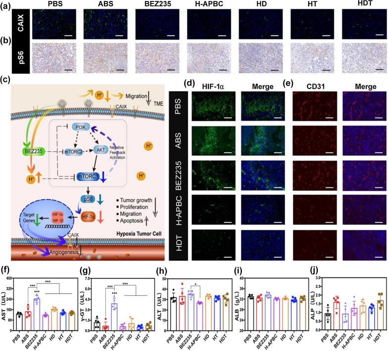
Fig. 5.
In vivo anti-hypoxia adaptation performance and biosafety assessment of H-APBC. Images of tumor tissues expressing CAIX (a) and pS6 (b) after different treatments. Scale bar: 80 μm (CAIX), 120 μm (pS6). c Anti-cancer strategy of H-APBC under hypoxic condition. Under hypoxia, H-APBC aggregated in tumor tissues, inhibited CAIX expression and released BEZ235 to attenuate TME acidosis, increase intracellular acidity, and exert the enhanced PI3K/AKT/mTOR pathway inhibition accompanied by decreased pS6 protein expression. H-APBC more strongly inhibited HIF-1α expression and mitigated metastasis and angiogenesis. Immunofluorescence images of expressed HIF-1α (d) and CD31 (e) in different groups. Scale bar: 80 μm (HIF-1α),120 μm (CD31). f–j Serum aspartate aminotransferase (AST), γ-glutamyltransferase (γ-GT), alanine aminotransferase (ALT), albumin (ALB) and alkaline phosphate (ALP) levels recorded for mice in the treatment groups. *p < 0.05, **p < 0.01, and ***p < 0.001

