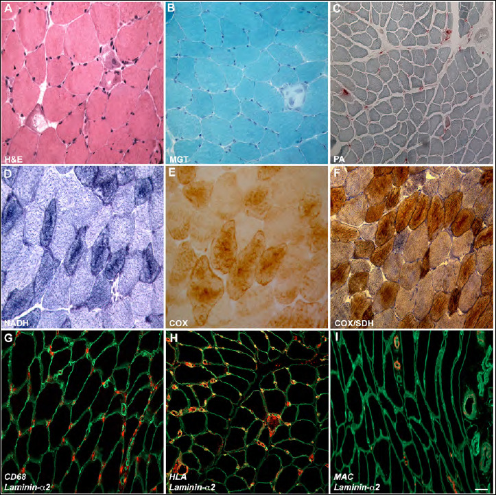
Figure 1.

Light microscopy observations. H&E (A) and MGT (B) show few degenerative and necrotic fibres and normal connective tissue (magnification 400 x). Acid phosphate reaction (C) is positive in necrotic fibres and at interstitial level (magnification 200 x). NADH (D) signal is increased in type I fibres, COX (E) and COX-SDH (F) reactions show COX signal reduction in some scattered fibres with normal SDH activity (magnification 400 x). Immunohistochemistry shows CD68 (macrophage) positivity (G), HLA positivity at membrane level and in the cytoplasm of necrotic fibres (H) and MAC positivity in capillaries (I). Laminin-α2 (green) was used for membrane counterstaining (magnification 400 x).
