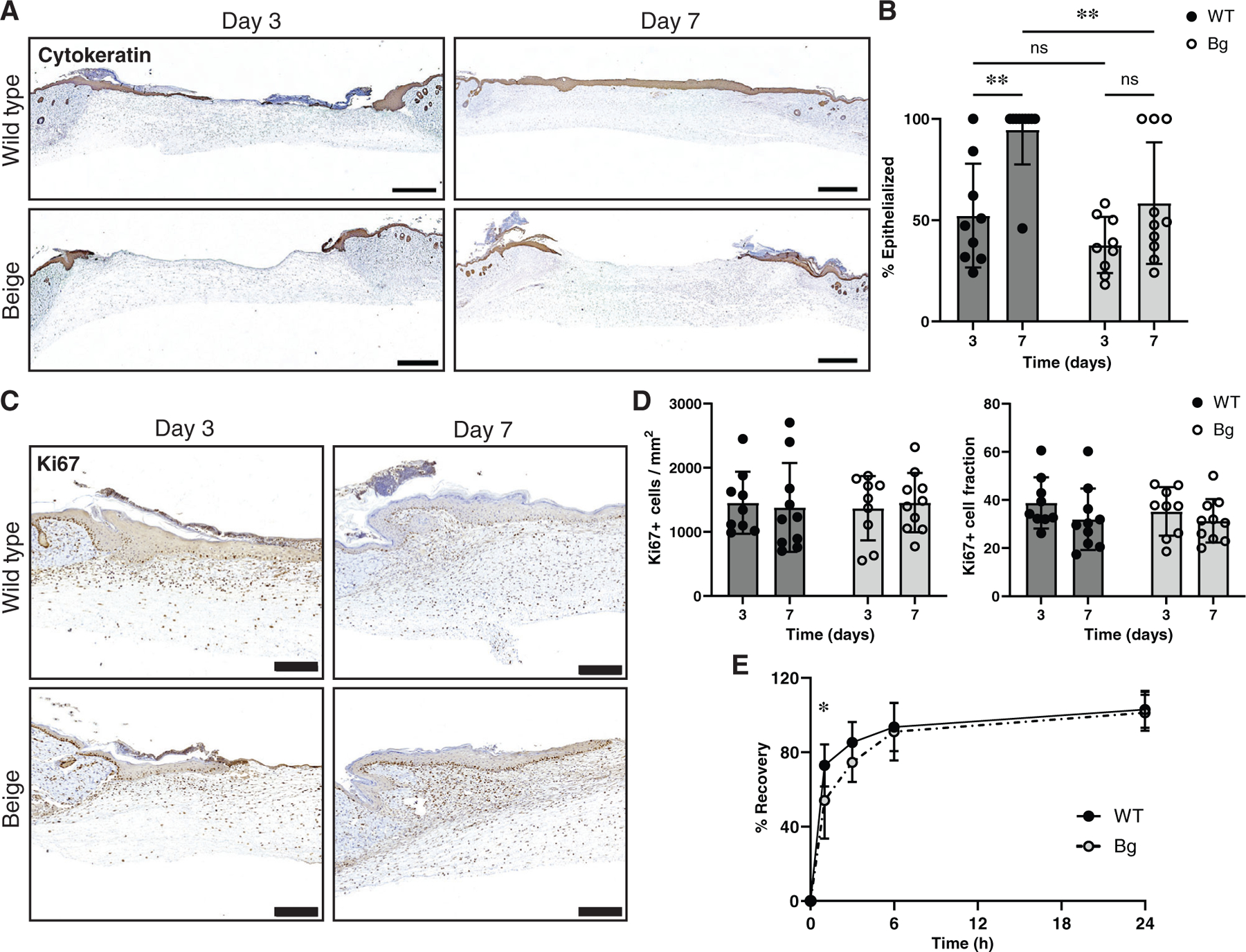
FIGURE 5.

Epithelialization, proliferation, and barrier function. (A) Tissue sections were stained with a pan-cytokeratin marker to examine the epithelialization of wounds between groups. Scale bar = 1 mm. (B) Measurements of epithelialization (cytokeratin positive cellular migration distance/full wound length) showed an increase in epithelialization in WT sections between days 3 and 7 (p = 0.0015) but not Bg (p = 0.2127). Furthermore, Bg wounds at day 7 were significantly less epithelialized compared to WT wounds at day 7 (p = 0.0058) (n = 9–10 per group). (C) Tissue stained with Ki67 were used to measure the proliferation of the epithelial layer. Scale bar = 200 μm. (D) There were no differences seen in either the number of Ki67 positive cells per area or the number of Ki67 positive cells per total cells (n = 9–10 per group). (E) Transepidermal water loss measurements taken showed that Bg mice had a delay in recovery of barrier function at 1-h post procedure (p = 0.0325) compared to WT mice. After 24 h, barrier function had been restored in both groups (n = 7–8 per group). *p ≤ 0.05, **p ≤ 0.01
