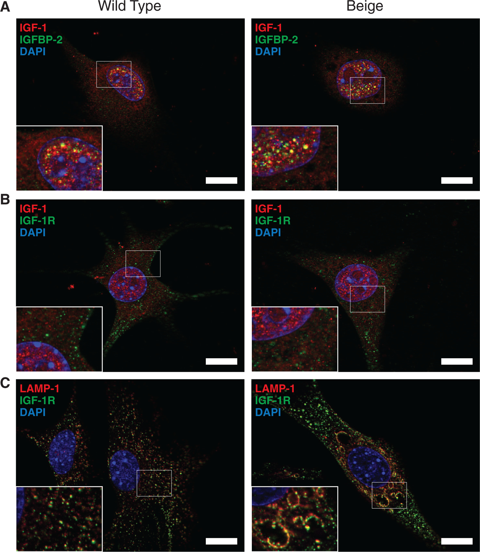
FIGURE 8.

Localization of IGF associated proteins in fibroblasts. (A) Co-stain of IGF-1 (red) and IGFBP-2 (green) shows nuclear colocalization (yellow) of the proteins based on the microscopic resolution (0.24 μm). Colocalization is not seen in the cytoplasm or in the nucleoli of the nucleus. (B) IGF-1 (red) and IGF-1 receptor (IGF-1R) (green) do not appear to colocalize. IGF-1R appears mostly cytoplasmic. (C) Lysosomes stained with LAMP-1 (red) and IGF-1R (green) show colocalization associated with the membrane of lysosomes. DAPI (blue) is used to visualise nuclei and void regions in nuclei are nucleoli. Scale bar = 20 μm
