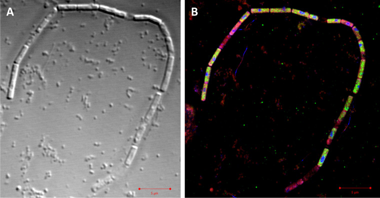
FIG 3.
Differential interference contrast micrograph (A) and superresolution structured illumination microscopy (SR-SIM) overlay image (B) of a filament assigned to Methanosaeta. Staining was achieved using Nile red (red) for lipids, probe Arch915 labeled with four 6-carboxyfluorescein molecules (green) for rRNA, and DAPI (blue) for DNA. Bar, 5 μm. For individual and dual overlay SR-SIM images, see Fig. S5.

