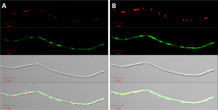
FIG 5.
LIVE/DEAD staining of a filament assigned to Methanosaeta in images with optimal exposure (A) and overexposure (B) to visualize regions of weak staining. In the DIC micrograph, dead (red) cells had less biovolume than live (green) cells. Overexposure showed a weak green staining of cells and short red cells separating green cells similar to spacer plugs. We assigned the short dead cells to unequally produced daughter cells that precede filament separation of Methanosaeta based on the observation of two spacer plugs between cells visible by Nile red staining (Fig. S8 and S9) and Methanosaeta microcells in thin-section TEM images (Fig. S11, S15, and S16). Bar, 5 μm.

