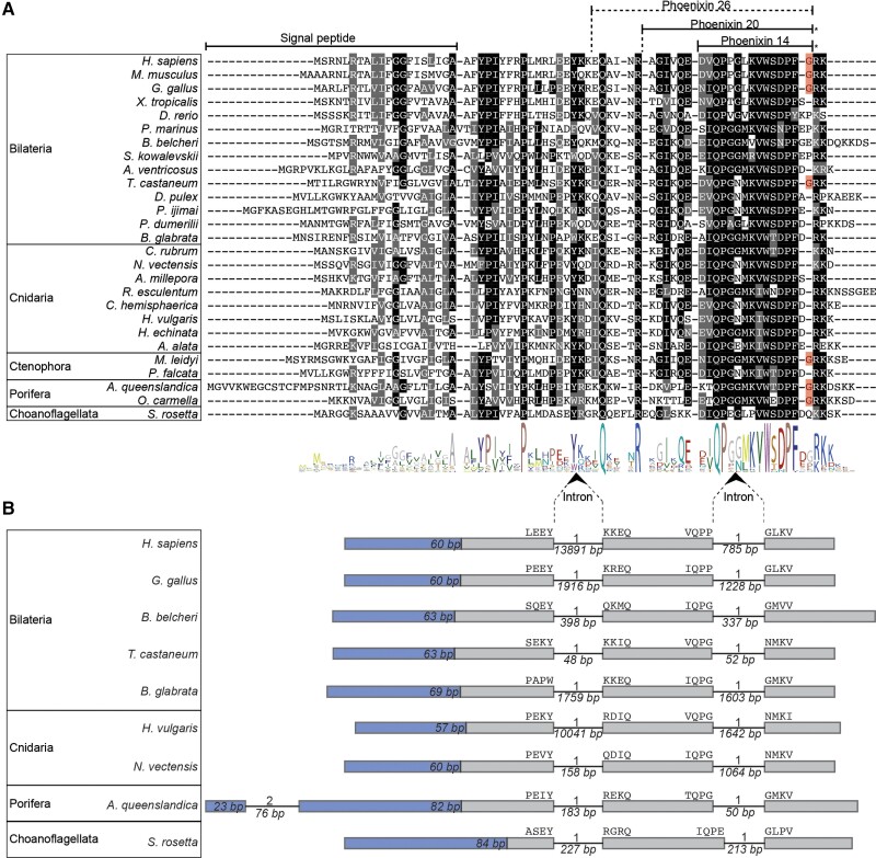
Fig. 1.
Sequence alignment and genomic structure of PNX precursors. (A) Alignment of the PNX precursors containing the PNX peptides. Predicted signal peptides and mature peptides are indicated with lines. Residues that are conserved in more than 50% of the sequences are shown in black, and conservative substitutions are shown in gray. Amidation sites are highlighted in red. (B) Exon–intron structure of PNX precursor genes. The regions encoding the signal peptides are in blue with their length indicated. Amino acids encoded at the exon–intron junctions are shown above the exon boxes. Introns are shown as lines, and their length in base pairs is indicated below. The intron phase is shown above the introns.

