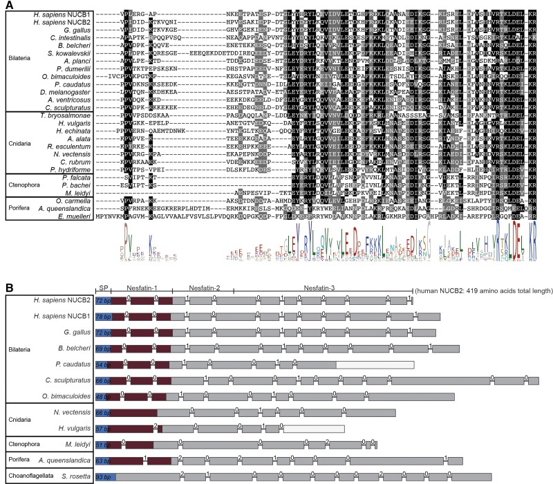
Fig. 2.
Sequence alignment of nesfatin-1 and genomic structure of NUCB precursors. (A) Alignment of the N-terminal NUCB precursor region containing the nesfatin-1 peptides. The conserved residues are highlighted, with conservation in more than 50% of sequences shown in black, and conservative substitutions shown in gray. (B) The genomic exon–intron structure of NUCB precursors. The regions encoding the signal peptides are shown in blue. The nesfatin-1-peptide coding region is indicated in dark red. Introns are shown as lines, with the phase of the introns shown above the lines. An empty/white box indicates a missing part in the mRNA-genome alignment.

