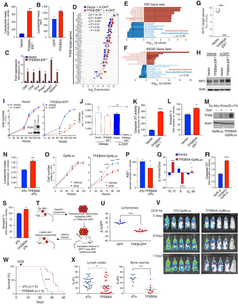
Figure 4.
TFEB functions as a tumor suppressor in Myc-driven lymphoma. A, Eμ-Myc lymphoma cells expressing vector or TFEBSA-ERT2 were treated with vehicle or 25 nmol/L 4-OHT for 4 days and analyzed for lysosomal mass by flow cytometry after staining cells with Lysotracker DND99. B, Eμ-Myc lymphoma cells were transduced for 48 hours with retroviruses expressing GFP or TFEBSA plus GFP, and GFP+ cells were analyzed for lysosomal mass. C and D, Eμ-Myc lymphoma cells expressing vector or TFEBSA-ERT2 were treated as in A, and then analyzed for the indicated TFEB target genes by qRT-PCR (C) or RNA-seq (D; n = 3 for both). For D, the log2 gene expression profile of TFEB target genes is shown as a dot plot that is ordered on the basis of expression; each dot represents one sample, and its size corresponds to its statistical significance as shown. E and F, GSEA of significant, differentially expressed genes from RNA-seq analysis were performed using the PID (E) and KEGG (F) databases. G, Box plot comparing the change of canonical MYC target genes (PC1) in Vector + 4-OHT versus TFEBSA-ERT2 + 4-OHT RNA-seq data. H, Immunoblot analysis of Myc protein levels in Eμ-Myc lymphoma cells expressing vector or TFEBSA-ERT2 and treated as in A. I–L, Eμ-Myc lymphoma cells expressing vector or TFEBSA-ERT2 were assessed for cell proliferation over 6 days (inset shows TFEBSA-ERT2 protein expression; I); colony-forming potential in methylcellulose (day 14; J); and apoptosis after 4 days of 4-OHT treatment, by measuring caspase-3/7 and Annexin V staining, respectively (I–L, n = 3). M–S, Western blot analysis of TFEBSA and actin protein levels in Eμ-Myc;Eμ-rtTA2 lymphoma cells expressing vector (GpNLuc or dTo) or TFEBSA (TFEBSA-GpNLuc or TFEBSA-dTo) 2 days after treatment with vehicle or Dox (M); lysosomal mass of these cells treated ± Dox (48 hours; N); cell numbers over 120 hours (O); cell proliferation index, as determined by Ki67 staining (48 hours; P); cell-cycle analysis with propidium iodide (48 hours; Q); and apoptosis (caspase-3/7 activity and Annexin V staining after 48 hours; R and S). T and U, Eμ-Myc lymphoma cells were transduced with retrovirus expressing GFP or GFP plus TFEBSA. T, A 50:50 mix of control or TFEB-expressing GFP+:GFPNeg Eμ-Myc lymphoma cells from each transduction was transplanted intravenously into congenic recipient mice (n = 8). U, Lymphomas arising in transplanted animals in S were assessed for percent of GFP+ lymphoma cells. V,In vivo imaging of Nod/Scid mice transplanted with Eμ-Myc;Eμ-rtTA2 lymphoma cells expressing vector (GpNLuc) or TFEBSA-GpNLuc at days 3, 6, and 7 after Dox chow was provided ad libitum. W, Survival of syngeneic mice transplanted with Eμ-Myc;Eμ-rtTA2 lymphoma cells expressing vector (dTo) or TFEBSA-dTo; Dox chow was provided at day 3 posttransplant (arrow). X, Percent dTo+ B220+ cells isolated from lymph nodes or BM of diseased recipient mice in V. Statistical analysis: A–C, I–L, N–S, U, and X: Student t tests were performed. W,χ2 test was performed. *, P ≤ 0.05; **, P ≤ 0.01; ***, P ≤ 0.001; ****, P ≤ 0.0001.

